Basics of Knee Aspiration
Case
A 70-year-old female with a past medical history of insulin-dependent diabetes mellitus, gout, COPD, chronic atrial fibrillation on apixaban, and obstructive sleep apnea complained of sudden-onset right knee pain with inability to bear weight during a hospital admission for syncope. Her knee joint had never before been affected by gout. Plain films and Commuted Tomography of her knee did not show any fracture or dislocation but revealed a moderate joint effusion. Arthrocentesis was performed to evaluate etiology of effusion and rule out septic joint.
Background
The foremost consideration in the presentation of joint pain is septic arthritis. Septic arthritis is classically characterized by red, hot, swollen joint with associated effusion and fever and limited passive and active range of motion. [1] The knee is implicated in 50% of septic joints. There is good reason to be concerned: patients who present with a new-onset swollen and painful joint have up to a 27% chance of having septic arthritis. Importantly, absence of fever or chills, labwork, and history do not reliably exclude septic joint. The most sensitive physical exam findings are pain with motion, tenderness, and joint effusion/swelling. [2] Joint pain and joint effusion may, in fact, be the only finding in septic arthritis [3], and findings commonly associated with septic joint such as fever and erythema are only moderately sensitive. [2] As a result, arthrocentesis is the most valuable test in the evaluation of a potential septic joint and is the required test to adequately assess and rule in or out a septic joint. Due to the necessity for diagnosis there exist no absolute contraindications to arthrocentesis. Relative contraindications include cellulitis, dermatitis, other lesion over the joint; coagulopathy; prosthetic joint; and difficulty identifying landmarks. [4]
Arthrocentesis is the insertion of a needle into a joint space and may involve aspiration (removing fluid from the joint) or injection (inserting fluid into the joint), or both. In the case of septic arthritis, joint aspiration is performed diagnostically to analyze synovial fluid. Additionally, joint aspiration may be therapeutic – joint effusions stretch the joint capsule and are often painful. During arthrocentesis, it is important to follow sterile technique.
Arthrocentesis is indicated in any case involving acute monoarthritis or acute nontraumatic effusion if there is diagnostic concern for a septic etiology.
Equipment
Obtain clean or sterile gloves, gauze, povidone iodine, chlorhexidine solution, or other antiseptic option, as well as a sterile skin marking pen. You will also need a hemostat, local anesthetic, several needles (27 gauge needle for anesthetic, and 18 gauge needle for fluid aspiration) and several syringes for fluid aspiration (10, 20, 30, 60 mL). Consider obtaining specimen and culture tubes (for cell count and differential, culture and gram stain, crystal analysis), bandaid (or other post procedure dressing).
Procedure
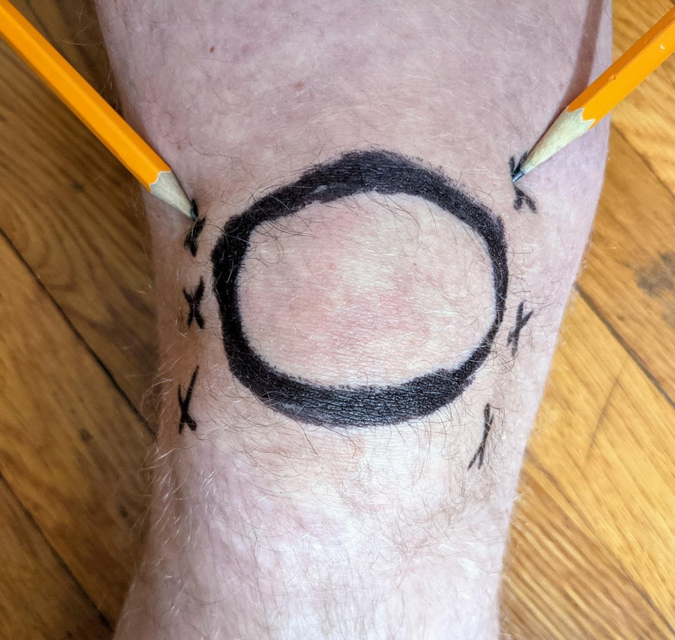
As with all procedures, obtain informed consent. Identify your landmarks and locate point of entry into joint space. Consider marking your access site with the sterile pen. As below the knee joint may be entered from parapatellar, suprapatellar, or infrapatellar approach.

Parapatellar approach (supine)
Suprapatellar approach (supine)

Infrapatellar approach (seated)
Ensure there is no debris or dirt on skin and apply povidone iodine or chlorhexidine, and wait for it to dry to ensure that the area is sterile. Apply 3-7 mL anesthetic (commonly 1% lidocaine) into subcutaneous tissues but not into the joint space using the 27 gauge needle.
Insert the 18 gauge needle attached to the 10 cc syringe into the joint space and aspirate fluid. If the needle meets bone, then withdraw the needle and re-advance at a different angle, aiming horizontally but deep to the patella. One can “milk” the joint by applying pressure to the joint effusion via a helper or your free hand, avoiding side of the joint with the needle insertion point. If the needle meets the patella, re-direct deep to patella.
If the syringe fills with synovial fluid, use a hemostat to hold the needle hub while removing the syringe. Put appropriate quantity of fluid into containers or tubes. Return the syringe with the needle still within the joint in order to withdraw additional fluid. This can be repeated multiple times.
If the syringe does not fill with fluid, i.e. the quantity of aspiratable fluid is less than the syringe volume, then remove the needle and syringe. Put appropriate quantity of fluid into containers or tubes. wipe off the antiseptic and put a dressing on the skin. Dispose of equipment and sharps appropriately. [3, 4]
Case conclusion
Arthrocentesis of patient’s joint 30 mL yellow synovial fluid. Analysis showed a nucleated cell count of 25,000, and calcium pyrophosphate crystals. While synovial fluid nucleated cell count is often cited at 50,000 as “cutoff” for consideration of septic joint, in reality this marker has been associated with summary estimate sensitivity of only 56 in Carpenter’s evidence-based review of diagnosis of adult septic arthritis. [1] In the case presented here, the finding of calcium pyrophosphate crystals in the fluid points toward crystal arthropathy, but it is important to remember that synovial fluid culture is the ultimate diagnostic for septic arthritis.
Patient was started on colchicine. NSAIDs were held due to cardiac history.
Take aways
A new-onset swollen and painful joint should prompt evaluation for septic arthritis.
Arthrocentesis is the most valuable test in evaluating for septic arthritis.
Three approaches may be used in knee arthrocentesis.
Synovial fluid WBC > 50,000 is reasonably specific but not particularly sensitive for septic joint; synovial fluid culture is ultimately diagnostic.
AUTHOR: Rachel Smith Shain, MD
FACULTY REVIEWER: Adam Aluisio, MD
References
[1] Goldenberg, Don L.; Sexton, Daniel J. Septic arthritis in adults. Uptodate.com. https://www.uptodate.com/contents/septic-arthritis-in-adults. Published July 2020. Accessed January 2021.
[2] Carpenter, Christopher R et al. “Evidence-based diagnostics: adult septic arthritis.” Academic emergency medicine : official journal of the Society for Academic Emergency Medicine vol. 18,8 (2011): 781-96. doi:10.1111/j.1553-2712.2011.01121.x
[3] Tintinalli, Judith, ed.. Tintinalli’s Emergency Medicine: A comprehensive Study Guide. Eighth Edition. United States of America. McGraw-Hill Education. 2016. pp. 1929-1933.
[4] Reichman, Eric, ed. Reichman’s Emergency Medicine Procedures. Third Edition. China. McGraw-Hill Educatin. 2019. pp. 819-841.