A Case of Near Syncope
CASE
A 69-year-old female with a history of hypertension presents to the ED with near syncope. She had been walking around Newport on a hot day with her family when she began to feel lightheaded. Her family noted that she appeared pale and diaphoretic. She did not lose consciousness but she continued to feel weak and was brought to the emergency department.
On arrival, she attributes her weakness and lightheadedness to heat exposure and denies any chest pain, abdominal pain, or respiratory symptoms. Notably, her oxygen saturation is 88% and respiratory rate is 24/min. These findings raise concern for pulmonary embolism, with dehydration/heat exposure and ACS also possible.
Her other vital signs include blood pressure of 110/70, HR 79, temperature 95.5, and glucose 131. Her EKG shows normal sinus rhythm and she has a normal chest X-ray. Her physical exam shows no asymmetric calf swelling, calf tenderness or palpable cord.
Initial labs are as follows:
CBC, BMP: all within normal limits
VBG: pH 7.46, pCO2 35 (42-50), pO2 49 (30-50)
Troponin I: 97 (2-15)
BNP: 311.8 (0-100)
Lactate: 2.4
D-dimer: 4,044
This patient’s respiratory alkalosis and elevated D-dimer in the setting of tachypnea and hypoxia are concerning for pulmonary embolism. A CT PE is performed which shows bilateral pulmonary emboli with overall moderate clot burden without evidence of right heart strain. There is a wedge-shaped ground glass attenuation in the left upper lobe, likely representing pulmonary infarct. The patient is started on heparin and admitted for further management.
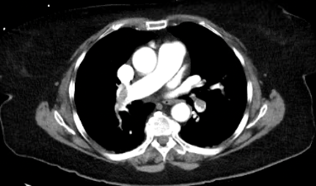
Figure 1. CTPE Findings
DISCUSSION
Right Heart Strain
If severe, an acute PE can cause pulmonary arterial hypertension and put significant strain on the right side of the heart. Right ventricular dysfunction is a predictor of poor outcomes in acute PE patients, so early detection is an important step in risk stratification and decision-making about thrombolysis or embolectomy.
On EKG, right ventricular dilatation can manifest as T-wave inversions in V1-4 and the inferior leads (III, aVF). Imaging with CT or echocardiogram can also be useful in assessing RV strain. CT is more sensitive than echocardiography in demonstrating right heart dysfunction, but both have specificity below 50%. On CT, there may be abnormal positioning of the interventricular septum, right ventricle enlargement, pulmonary trunk enlargement, and proximal venous signs (hepatic or azygous dilation, IVC reflux). On echocardiography, features of RV dysfunction include enlargement of the right ventricle or atrium, elevated RV pressures, tricuspid regurgitation and RV free wall hypokinesis.
Biomarkers and PE Prognosis
While echocardiography has been proven to be a helpful tool in evaluating right heart strain in patients with acute PE, it is associated with high cost and limited availability, and visualization of the right ventricle may be difficult in some patients. Therefore, cardiac biomarkers such as troponin I and brain/B-type natriuretic peptide (BNP) have been used for risk stratification. Cardiac troponins are sensitive indicators of cardiomyocyte damage and correlate well with the extent of right ventricular dysfunction in acute PE, possibly due to coronary artery compression causing ischemia in the setting of increased RV wall tension. BNP production in ventricular myocytes is stimulated by stretch, and studies have shown that it is also associated with RV dysfunction in PE. Importantly, both troponin and BNP can be increased as a result of other pathologies and should therefore be used cautiously.
Studies have also shown that the BNP/troponin ratio may be the best metric to use. BNP elevation out of proportion to troponin elevation is correlated with RV dilation and hypokinesis on echocardiogram. Patients who had lower ratios were less likely to require ICU admission and more likely to survive to discharge, 30 days, and 90 days.
Management
In hemodynamically stable patients, anticoagulation is generally indicated. However, the benefits of this therapy should be weighed with bleeding risks and an IVC filter is an acceptable option for patients at high risk. In some hemodynamically stable patients, thrombolysis or embolectomy may be indicated if there is a large clot burden, severe RV dysfunction, or other concerning features. Hemodynamically unstable patients should receive thrombolytic therapy. Surgical or catheter-based embolectomy should be performed in patients for whom thrombolysis is contraindicated or unsuccessful.
Author: Katherine Barry is a fourth year medical student at the Warren Alpert Medical School of Brown University.
Faculty Reviewer: Kristina McAteer, MD is an attending physician at Rhode Island Hospital and Newport Hospital.
REFERENCES
Bondarsky, E. Brain Natriuretic Peptide/Troponin I Ratio in Pulmonary Embolism. CHEST Journal, journal.chestnet.org/article/S0012-3692(17)32591-6/fulltext.
Burns, E, and Buttner, R. “Right Ventricular Strain.” Life in the Fast Lane • LITFL, 8 Mar. 2021, litfl.com/right-ventricular-strain-ecg-library/.
Kucher, N, Goldhaber, S., et al. “Cardiac Biomarkers for Risk Stratification of Patients With Acute Pulmonary Embolism.” Circulation, 4 Nov. 2003, www.ahajournals.org/doi/full/10.1161/01.CIR.0000100687.99687.CE.
“Treatment, Prognosis and Follow-up of Acute Pulmonary Embolism in Adults.” UpToDate, www.uptodate.com/contents/treatment-prognosis-and-follow-up-of-acute-pulmonary-embolism-in-adults.
Weerakkody, Y. “Right Heart Strain: Radiology Reference Article.” Radiopaedia Blog RSS, radiopaedia.org/articles/right-heart-strain?lang=us.