Yank that Ank! A Maisonneuve Fracture With An Associated Ankle Dislocation and a Discussion of the Identification, Management, and Disposition of Various Ankle Fractures in the Emergency Department.

CASE
A 24-year-old healthy male presented to the emergency department (ED) with right ankle pain. He was playing in a rugby game and was tackled by another player who landed on his right ankle. He was unable to bear weight or walk. No numbness, tingling or weakness. No other complaints.
On exam, there was an obvious deformity to the right ankle with some associated swelling (Figure 1). Skin was intact. The patient had tenderness to palpation of the ankle, proximal fibula, and navicular bone. He was neurovascularly intact, and compartments were soft.
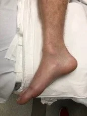
Figure 1. Right ankle with obvious deformity.
X-rays of the right foot, ankle, tibia/fibula, and knee demonstrated a lateral and posterior talar dislocation with medial malleolus avulsion fracture, segmented proximal fibular fracture, and a nondisplaced intra-articular navicular fracture (Figure 2) (Note: foot imaging not shown).

Figure 2. Radiographs showing talar dislocation, medial malleolar fracture, proximal fibular fracture.
DIAGNOSIS
This patient presented with an obviously dislocated ankle, and X-ray imaging also revealed a proximal fibular fracture consistent with a Maisonneuve (ankle + proximal fibula) fracture. In this case, it was clear that orthopedic surgery (ortho) consultation was indicated.
DISCUSSION
But what about the patients that come in with less dramatic fractures? What kinds of fractures need ortho consults in the ED? What kinds of fractures need to be admitted? What kinds of fractures can we splint by ourselves and send home?
This blogpost seeks to answer these questions by giving a brief overview of the management of different types of ankle fractures. A few disclaimers: the scope of this blogpost is limited to the management of ankle fractures in the ED. Additionally, the indications to consult ortho in this blogpost are influenced by the current practice at a tertiary care academic center. Guidelines and protocols regarding orthopedic consultation may vary in different regions and/or clinical settings.
Ankle Anatomy:
The ankle is made up of three bones: the tibia, fibula, and talus. The tibia and talus bear most of an individual’s weight. The fibula plays a small role in weightbearing, but it is critically important for the stability of the ankle joint. There are three groups of ligaments that hold these bones together and maintain stability in the anterior-posterior, medial, and lateral planes (Figure 3). [1] Ankle injuries can damage both bones and ligaments and can threaten the stability of the ankle joint. Patients with unstable fractures are at high risk of developing traumatic osteoarthritis, and therefore generally require surgical intervention. [2]

Figure 3. Ankle joint anatomy. [1]
EXAMINATION TIPS

When examining an ankle injury, it is important to check for punctures, lacerations, and bleeding. Any fracture with an overlying wound should be assumed to be an open fracture until proven otherwise (needs admission and urgent/emergent surgery). Be sure to examine the foot (particularly the navicular and 5th metatarsal) as well as the proximal fibula and knee. Don’t miss compartment syndrome: palpate all compartments in the lower leg and check for pain with passive stretch (may be limited due to competing pain from a broken ankle). [3]
IMAGING
Refer to the Ottawa Ankle Rules (OAR) when deciding which ankle injuries need imaging. [4] Order at least 3 views: Anterior Posterior (AP), Mortise (AP but with the foot internally rotated by about 10 degrees) (figure 4), and Lateral (figure 5). Imaging should extend proximal and distal to the injury to assess for other fractures. If initial X-rays are equivocal, consider imaging the opposite ankle for comparison and/or ordering stress films (in which the ankle joint is stressed to evaluate for medial clear space widening which would indicate deltoid ligament injury and joint instability). [5] Consider advanced imaging (CT, MRI) for extreme mechanisms of injury such as crush injuries or major axial loads. [3]

Figure 4. AP and Mortise views. [5]

Figure 5. Approach to AP, Mortise, and Lateral views. [6]
ISOLATED DISTAL FIBULA FRACTURES
The Danis-Weber classification system is used to describe isolated distal fibula fractures (figure 6).

Figure 6. Weber A, B, and C fractures. [2]
Weber A is a fracture of the fibula distal to the syndesmosis (where the ligaments hold the ankle bones together). Weber A fractures are generally of little clinical significance because the stability of the ankle joint is maintained. These fractures can be placed in a CAM boot or splinted and discharged with a referral for outpatient ortho follow up. [2]
Weber B fractures are fractures of the fibula at the level of the syndesmosis and can behave like a Weber A or a Weber C fracture. Stress imaging is indicated to evaluate for joint instability. If stress x-rays (particularly the mortise view) show medial clear space widening which is indicative of a rupture of the deltoid ligament, then the fracture is unstable and will likely benefit from orthopedic surgery. If no clear space widening is visualized, then the fracture is more likely to be stable and surgery may not be indicated. Depending on local practice patterns, Weber B fractures may be managed solely by the emergency physician by obtaining stress imaging, applying a splint, and discharging with outpatient ortho follow up, or they may warrant an ortho evaluation in the ED. [2]
Weber C fractures are fibular fractures proximal to the syndesmosis and are almost always unstable (figure 7). These fractures warrant ortho consultation in the ED. [8]

Figure 7. Weber C fracture. [7]
ISOLATED MEDIAL MALLEOLAR FRACTURE
Medial malleolar fractures are frequently associated with other fractures, so X-rays should be carefully examined for other injuries. If they are truly isolated and stable, they may be splinted and discharged with ortho follow up. [2]
ISOLATED POSTERIOR MALLEOLAR FRACTURE
These are rarely (< 5% of the time) seen without other associated fractures [9]. Because of this ortho consultation should be strongly considered. If they are truly isolated (figure 8), and stable, they may be splinted and discharged with ortho follow up.

Figure 8. Posterior malleolar fractures. [6]
BIMALLEOLAR
Two malleoli are fractured; these are usually unstable and may require surgery. Consult ortho. [3]
TRIMALLEOLAR
Three malleoli are fractured, (Figure 9); these are unstable. Consult ortho. [3]

Figure 9. Trimalleolar fracture. [6]
PILON FRACTURE
French for “piledriver,” this fracture is frequently caused by a downward force pushing the tibia and fibula into the talus. The tibia and fibula are usually comminuted (figure 10). Consult ortho. [10]

Figure 10. Pilon fracture. [10]
MAISONNEUVE FRACTURE
This is an ankle fracture with a syndesmotic injury and proximal tibial fracture (figure 11); unstable. Consult ortho. [1]

Figure 11. Maissoneuve fractures. [1]
ANKLE FRACTURE/DISLOCATIONS
These should all be urgently reduced (Figure 12). If there is any concern for neurovascular compromise, these should be reduced IMMEDIATELY (don’t even wait for X-rays). Consult ortho. [11]

Figure 12. Ankle fracture with dislocation. [ 11 ]
CASE RESOLUTION
Ortho was consulted and successfully reduced the patient’s ankle dislocation under conscious sedation. Post-procedure, the patient’s pain was greatly improved, and he was neurovascularly intact. Ortho splinted his ankle, and post-reduction X-rays were deemed adequate (Figure 13). The patient was discharged with follow-up for outpatient surgical management.

Figure 13. Post reduction and splinting X-rays. Note that there is still some medial clear space widening. This injury will require surgical management.
TAKE-AWAYS
When evaluating ankle injuries, don’t forget to examine the proximal fibula and foot and to image above and below the injury.
If fracture is stable, CAM boot or splint and outpatient ortho follow-up.
If fracture is unstable, consult ortho in the ED. Patients will need prompt surgical intervention. Depending on the injury and shared-decision-making between ortho and the patient, disposition could be admission vs. splint, non-weight-bearing, and prompt outpatient surgery.
If fracture is open, consult ortho (will require admission and urgent surgical intervention).
If fracture is dislocated, reduce it promptly and consult ortho.
If fracture is dislocated and there are concerns for neurovascular compromise, DO NOT WAIT for X-rays, DO NOT PASS GO, YANK THAT ANK! Then consult ortho.
AUTHOR: Gabe Padilla, MD is a first-year resident at Brown University/Rhode Island Hospital.
FACULTY REVIEWER: Michelle Myles, MD, is an assistant professor and clinician educator at Brown Emergency Medicine.
References:
1. Van Heest T, Lafferty P. Injuries to the Ankle Syndesmosis. J Bone Joint Surg Am. 2014;96:603-13.
2. Canton G, Sborgia A, Maritan G, et al. Fibula fractures management. World J Orthop. 2021;12(5): 254-269.
3. Wedmore, I, Young S, Franklin J. Emergency Department Evaluation and Management of Foot and Ankle Pain. Emerg Med Clin N Am. 2015;33:363–396.
4. Stiell I, Greenberg G, McKnight D, et al. Decision Rules for the Use of Radiography in Acute Ankle Injuries Refinement and Prospective Validation. JAMA. 1993;269(9):1127-1132.
5. Kellett J, Lovell G, Eriksen D, Sampson M. Diagnostic imaging of ankle syndesmosis injuries: A general review. Journal of Medical Imaging and Radiation Oncology. 2018;62:159-168.
6. Yu J, Cody M. A template approach for detecting fractures in adults sustaining low-energy ankle trauma. Emerg Radiol. 2009;16:309–318.
7. Gougoulias N, Sakellariou A. When is a simple fracture of the lateral malleolus not so simple? Bone Joint J. 2017;99-B:851-855.
8. Aiyer A, Zachwieja E, Lawrie C, Kaplan J. Management of Isolated Lateral Malleolus Fractures. J Am Acad Orthop Surg 2019;27:50-59.
9. Odak S, Ahluwalia R, Unnikrishnan P, Hennessy M, Platt S. Management of Posterior Malleolar Fractures: A Systematic Review. The Journal of Foot & Ankle Surgery. 2016;55:140–145.
10. Wei S, Han F, Lan S, Cai X. Surgical treatment of pilon fracture based on ankle position at the time of injury/initial direction of fracture displacement: A prospective cohort study. International Journal of Surgery. 2014;12:418-425.
11. Goost H, Wimmer M, Barg A, Kabir K. Fractures of the Ankle Joint. Investigation and Treatment Options. Dtsch Arztebl Int. 2014; 111:377−88.