Shoes matter! LE Degloving injury following a MCC

Case
63 y.o. male arrived to the ED following a motorcycle crash (MCC) in which he was riding at an unknown speed when he suddenly lost control and was launched off of his motorcycle. He was wearing a helmet, and he endorses multiple injuries throughout his body including his right arm, bilateral legs, chest, and abdomen. He denies any loss of consciousness (LOC) or head strike. He arrived via EMS as a level A trauma to the emergency department.
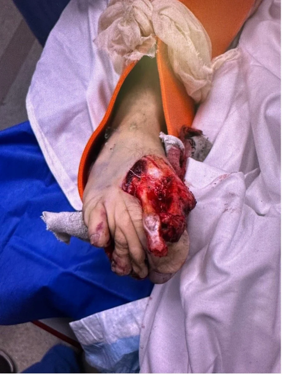
Per EMS, his vitals had been stable prior to arrival. Primary survey was intact, with dopplerable dorsalis pedal pulses and palpable femoral pulses on the left, none present on the right. Secondary Survey was notable for visible deformity of the right arm, as well as laceration to the lateral right hand with an obvious open fracture to the third digit. He also had a visible open fracture to the right shin, open fracture of the right ankle and right foot, and degloving injury to the toes of the right foot. Other notable injuries were road rash and abrasions present in the abdomen, chest wall, and left upper and lower extremities. He had chest wall tenderness with palpable crepitus, however there was no pneumothorax or subcutaneous air visible on chest x-ray imaging.
A CT angiography was obtained, which showed a bifrontal subarachnoid hemorrhage, numerous rib fractures, and numerous comminuted and displaced lower extremity fractures. Threatened limb protocol was activated, and the patient was emergently taken to the operating room for a below-the-knee amputation of the right lower extremity.
Diagnosis
This patient suffered severe soft tissue damage with an open degloving injury, along with multiple long bone fractures, an exposed tibial fracture, forefoot and midfoot fractures including fractures of the second through fifth metatarsals, cuboid, and multiple phalanges.
Discussion
Trauma to the extremities requires multi-focal approach considering the nerves, bones, vessels, and soft tissue to evaluate for the likelihood of “mangled extremity”. In most instances, saving a limb that would otherwise be lost without intervention can be attempted even if the patient has a mangled extremity. However, at times, the injury to the lower extremity is so severe that primary amputation at the initial operation is required to save the patient’s life. According to the civilian National Trauma Data Bank, traumatic injury results in an estimated 3700 major amputations annually[1].
Worth considering during the decision making path is that the presence of open fracture significantly increases the risk of osteomyelitis and limb loss depending upon the severity of injury to the associated soft tissues[2]. Use of the MESS (Mangled Extremity Severity Score) is a rating scale for lower extremity trauma, based on skeletal, soft-tissue damage, limb ischemia, shock, and age[3]. A retrospective survey and a prospective trial assessed the accuracy of MESS for limb evaluation and found that a MESS value greater than or equal to 7 predicted amputation with 100% accuracy. The application lies in its usefulness triaging trauma patients whose lower extremity injuries warrant primary amputation.
While the MESS score has its merits in evaluating the severity of a lower extremity injury, the initial resuscitation, diagnostic evaluation, and management of the trauma patient is based upon protocols from the Trauma and Life Support program (ATLS)[4]. Control of hemorrhage by either direct pressure or tourniquet remain the primary methods of controlling blood loss; however, depending on whether the patient may require transport or manipulation during management other methods such as endovascular occlusion devices, topical agents, or external fixation clamps may be used[5].
An adequate primary survey of the patient ought to include evaluation of the extremity, assessing the four functional elements: nerves, vessels, bones, and soft tissue. In this awake (and cooperative) patient, it was easy to assess for peripheral nerve damage in the form of motor or sensory deficit (both were absent in this case). If the patient were unconscious or uncooperative, then gross asymmetry, deformity, or lack of movement can serve as an evaluation. It is worth noting that a repeat evaluation is necessary to assess for changes in the patient’s neurovascular status.
This patient had an open degloving injury of the 5 digits that began in the mid-foot. Degloving injuries are categorized as pure degloving (involving only the skin, without futher bone or soft tissue involvement)[1] and further classified as open or closed, where a closed injury represents a traumatic separation of the skin and subcutaneous tissues from the underlying fascia without a break in the skin[2].
The severe fractures and soft tissue injuries in this patient made it impossible to assess for a dorsalis pedis pulse (DP). The soft tissue of the calf and ankle were not as impacted; however, the posterior tibialis (PT) was absent on palpation and by Doppler evaluation, moreover, the foot was cool to touch - all hard signs of arterial injury[3].
Commonly, vascular injury can be evaluated with the use of an Ankle-Brachial Index (ABI), an Injured Extremity Index (IEI), or Arteriography. The IEI is performed to assess for occult vascular injury, as a normal IEI (>0.9) has a high negative predictive value for vascular injury,[4] however this can be omitted in patients with hard signs arterial injury such as this one. Arteriography via CT imaging may be necessary to exclude vascular injury in hemodynamically stable patients, though in this case imaging was obtained not to rule out vascular injury but rather to better assess the extent of the osseous and neurovascular injury prior to operative management.
The long bone and tibial-fibula fractures present high risk of damage to the femoral, sciatic, and deep peroneal nerves as well as the aforementioned vascular injuries.
The immediacy of management in the emergency department and adequate triaging lies in the risk associated with severe or extensive muscle tissue damage such as rhabdomyolysis, ischemia of reperfusion, osteomyelitis, and its possible complications.
This case required collaboration between the Emergency Department and the Vascular Surgery and Orthopedic Surgery teams for operative management and close follow-up. In rural areas or situations with limited resources it is important for Emergency Physicians to be able to correctly assess the extent of the damage and triage the injuries that most require early amputation.
Case Resolution
This patient received an initial below-the-knee amputation of the right lower extremity, with a consequent second surgery requiring collaboration of the Orthopedic and Plastic surgery teams to close the skin flap and prep the wound for eventual outpatient evaluation for fitting of a prosthetic limb.
Author: Axel Schlossberg, MD, is a second year emergency medicine resident at Brown University/Rhode Island Hospital.
Faculty Reviewer: Kristina McAteer, MD, is an attending emergency medicine physician at Rhode Island Hospital and Newport Hospital.
References:
1. Dillingham TR, Pezzin LE, MacKenzie EJ. Incidence, acute care length of stay, and discharge to rehabilitation of traumatic amputee patients: an epidemiologic study. Arch Phys Med Rehabil. 1998 Mar;79(3):279-87. doi: 10.1016/s0003-9993(98)90007-7. PMID: 9523779.
2. Meling T, Harboe K, Søreide K. Incidence of traumatic long-bone fractures requiring in-hospital management: a prospective age- and gender-specific analysis of 4890 fractures. Injury. 2009 Nov;40(11):1212-9. doi: 10.1016/j.injury.2009.06.003. Epub 2009 Jul 5. PMID: 19580968.
3. J Trauma. 1990 May;30(5):568-72; discussion 572-3. Doi: 10.1097/00005373-1990050000-00007. PMID: 2342140
4. American College of Surgeons Committee on Trauma. Advanced Trauma Life Support (ATLS) Student Course Manual, 9th ed, American College of Surgeons, Chicago 2012.
5. Bulger EM, Snyder D, Schoelles K, Gotschall C, Dawson D, Lang E, Sanddal ND, Butler FK, Fallat M, Taillac P, White L, Salomone JP, Seifarth W, Betzner MJ, Johannigman J, McSwain N Jr. An evidence-based prehospital guideline for external hemorrhage control: American College of Surgeons Committee on Trauma. Prehosp Emerg Care. 2014 Apr-Jun;18(2):163-73. doi: 10.3109/10903127.2014.896962. PMID: 24641269.
6. Yan H, Gao W, Li Z, Wang C, Liu S, Zhang F, Fan C. The management of degloving injury of lower extremities: technical refinement and classification. J Trauma Acute Care Surg. 2013 Feb;74(2):604-10. doi: 10.1097/TA.0b013e31827d5e00. PMID: 23354258.
7. Nickerson TP, Zielinski MD, Jenkins DH, Schiller HJ. The Mayo Clinic experience with Morel-Lavallée lesions: establishment of a practice management guideline. J Trauma Acute Care Surg. 2014 Feb;76(2):493-7. doi: 10.1097/TA.0000000000000111. PMID: 24458056.
8. Nance ML. National Trauma Data Bank Annual Report. 2012. http://www.facs.org/trauma/ntdb/pdf/ntdb-annual-report-2012.pdf (Accessed on October 22, 2013).
9. Lynch K, Johansen K. Can Doppler pressure measurement replace "exclusion" arteriography in the diagnosis of occult extremity arterial trauma? Ann Surg. 1991 Dec;214(6):737-41. doi: 10.1097/00000658-199112000-00016. PMID: 1741655; PMCID: PMC1358501.