Ruby’s POCUS Pearls: Kidney Ultrasound
“…You might see a ‘twinkle’ artifact indicating a kidney stone”
Introduction Video
Kidney Stones (Hydronephrosis) 5MS (2024) | Videos & Movies on Vimeo
Pearls & Pitfalls
Scan in BOTH longitudinal and transverse planes.
To avoid rib shadows, oblique the probe to align with the rib space rather than holding the probe straight up and down.
Slowly fan through the kidney in its entirety to evaluate for hydronephrosis and for the presence of a kidney stone.
Hydronephrosis appears anechoic (black) and will spread from the renal pelvis towards the calyces and cortex depending on the grade (Image 1). Do not confuse it with a cyst (Image 2).
Renal vasculature may appear prominent (and also anechoic) leading you to suspect there is hydronephrosis. Use color Doppler to help differentiate hydronephrosis from renal vasculature. Hydronephrosis will not have color flow.
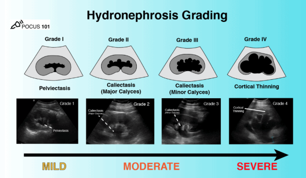
Image 1: Hydronephrosis Grading (mild involves pelvis, moderate involves calyces, severe involves cortex)
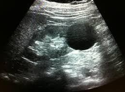
Image 2: Kidney Cyst. Anechoic (black) structure is in the periphery (cortex), not in the renal pelvis. Simple cysts are round and anechoic without internal septations
Kidney stones appear as focal echogenic areas (bright white) with posterior shadowing.
When suspecting a kidney stone, you can place the color Doppler box over the area and you might see a "twinkle" artifact indicating a kidney stone (Image 3).
Remember to also fan through the bladder in both planes and interrogate the posterior wall of the bladder for possible ureterovesicular junction (UVJ) stone (Image 3).
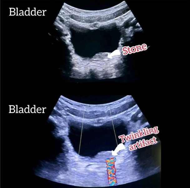
Image 3: Twinkle artifact occurs when color doppler is placed over a stone
To assess the ureteral jets, the bladder should be full and visualized in the transverse plane (Image 4).
Place color doppler box on the posterior wall of the bladder (furthest from probe or from top of screen) at the area of the trigone.
Keep the probe still and try to visualize both right and left jets by watching for bursts of color shooting upwards from the posterior wall of the bladder.
The jets intermittently burst every 10-20 seconds, however in some cases can take longer.
The visualization of jets can reassure against obstruction, however if they are not visualized you may have had the color box in the wrong place or not have been patient enough.
Pro tips for best documenting jets:
When the color box is on, adjust the gain to just below the "noise threshold". Turn up the gain until you see noise (eg artifactual color doppler) then reduce right to the point the noise is gone. This is to be sure your color gain is at the appropriate level.
Once you have seen the color burst from the jet(s), immediately hit the freeze button and use the "cine" tab to rewind back to the moment the jet stream was visualized and then hit the store button to save. This will help with capturing them as they can burst up quickly
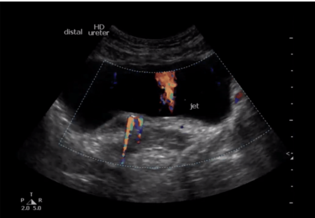
Image 4: Stone in the UVJ (on the left of the screen you see twinkle artifact from the UVJ stone and on the right you see ureteral jets, indicating no obstruction on the right side of screen)
POCUS for Colic Quality Project!
The POCUS division, Quality team and Urology are pleased to present our “POCUS for Colic” diagnostic algorithm for adults presenting to the ED with suspected renal colic (Image 5).
Our primary goal is to decrease unnecessary CT imaging for appropriate patients. Currently, our CT utilization for renal colic is nearly 50% higher than the national average. A reduction in CT for renal colic may improve department flow and decrease patient radiation.
To track our progress, CT utilization rates for patients diagnosed with nephrolithiasis will be included in your monthly provider metrics dashboard for your awareness and reference only.
This metric will not impact incentive compensation!
Of course, always get the imaging you feel is most appropriate for your patient, or if you aren’t sure how to interpret your POCUS.
If you need any help scanning you can sign up for scan shifts with Ruby: Wednesday to Friday between 7:30a-3:00p (rubypocustech@gmail.com), text at 603-781-1427 or secure chat on shift if you want to scan together. For any QPATH or Machine troubles please reach out for further assistance.
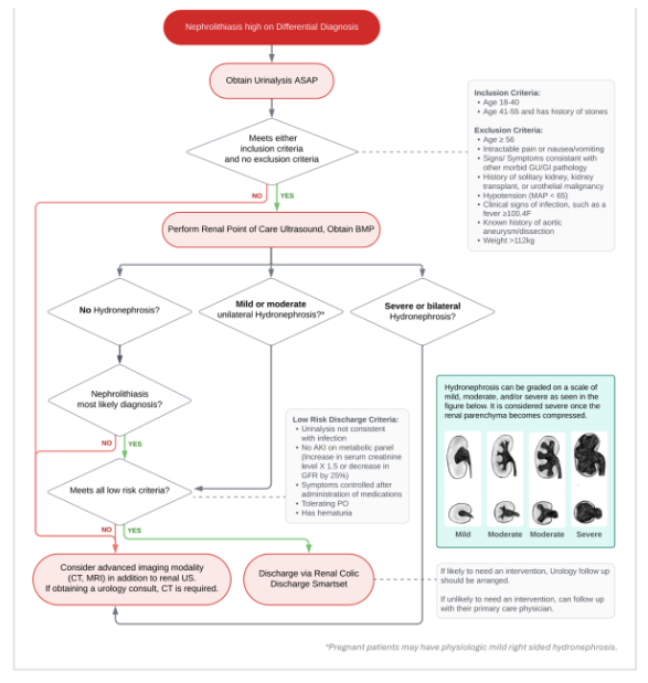
Image 5: Renal colic algorithm
Author
Ruby Agenor is an Ultrasound Clinical Specialist and ED Sonographer Educator at Brown Emergency Medicine.