Wait, you want to admit that clavicular fracture?
Case:
A 23-year-old male presents for a second opinion after a clavicle fracture one week prior. He was tackled in a softball game after hitting a player with a baseball. He had pain and deformity of his left mid-clavicle and initially presented to another hospital where imaging demonstrated a displaced mid-clavicular fracture. He was placed in a sling and referred for outpatient Orthopedic evaluation. He returned to the ED for ongoing pain and was found to have skin tenting (Figure 1). Repeat X-rays (Figure 2) showed “a segmental fracture of the mid left clavicular shaft with inferior displacement by 1.5 shaft widths.”
What is the next step in the management of this injury?

Figure 1: Physical exam showing displaced mid-clavicular fracture with skin tenting
Figure 2: Clavicle radiograph showing mid-shaft clavicle fracture with displacement
(Brief) Review of Clavicle Fractures:
o 2-5% of fractures in adults, 10-15% in children
o Bimodal distribution, males <30 yrs and elderly >70 yrs
o Mechanism: Fall on outstretched hand (FOOSH), direct fall on shoulder, direct trauma to shoulder (figure 3)
o Many classification systems (Craig, Neer, Edinburgh), none are particularly prognostic
o Generally divided anatomically: Medial-third, middle-third, lateral-third

Figure 3: Common mechanisms of injury for clavicle fracture
Management of Clavicle Fractures:
A. Medial-third fractures
a. Nearly always non-operative
b. Rarely displaced
c. Treatment: sling for comfort, early mobilization, non-union rates 4-8%
d. Surgical indications: Fracture displacement threatening mediastinal structures, soft tissue compromise
e. Children/adolescents: Differentiate physeal separation from sternoclavicular separation with CT as treatment differs
B. Middle-third fractures
a. Most common, greater than 2/3 of clavicle fractures
b. Historically non-operative, 1960’s studies showed 3x increase in non-union with surgical fixation
c. Changing dogma: Non-union now 10-15% in non-operative management
d. Newer research supports operative fixation: Shorter time to radiographic healing, less chance of non-union, higher satisfaction
e. Surgical indications: Young, active patients, displacement >100%, clavicle shortening >2 cm, significant deformity/skin tenting, multi-trauma. New studies: Non-union risk reduction of 87% with surgery
f. Children/adolescents: Majority non-operative (increased remodeling potential in children) UNLESS skin tenting or NV compromise. Sling with return to sports after radiographic evidence of healing (6-8 weeks)
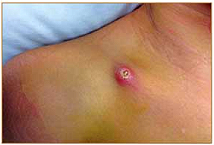
Figure 4: Skin tenting leading to erosion of overlying soft tissues
C. Lateral-third fractures
a. Treatment depends on specific fracture type, displacement, stability
b. Treatment: Sling, early mobilization
c. Surgical indications: Soft tissue compromise, medial fragment displacement (non-union ~30%)
d. Complications higher with delayed surgical fixation

Figure 5: Proportion of clavicular fractures
Take-home Points:
o Anticipatory guidance for patients
o Medial or lateral clavicle fractures; non-displaced, no soft tissue compromise: Generally non-operative with a sling for 6-8 weeks
o Middle third fractures that are displaced >100% the clavicle width, shortened, or a young active patient: Generally delayed operative intervention
o Soft tissue compromise (skin tenting), neurovascular compromise, mediastinal risk: Likely admission for urgent repair
o Calling Orthopedics: Classify anatomic location, open or closed
o Red flags for admission/urgent repair: Skin tenting, neurovascular compromise, open fracture, or mediastinal structures compromised
Case Outcome:
Orthopedic surgery was consulted. They admitted the patient for urgent operative intervention because of skin tenting and concern for skin breakdown and conversion to an open fracture. Overnight, the patient was checked regularly for skin breakdown around the tented skin. He was taken to the operating room the following morning for open reduction and internal fixation of the fracture.
Faculty Reviewer: Dr. Feden
Citations:
[1] Altamimi, S. A., and M. D. Mckee. "Nonoperative Treatment Compared with Plate Fixation of Displaced Midshaft Clavicular Fractures: Surgical Technique." JBJS Essential Surgical Techniques Os-90.Supplement_2_Part_1 (2008): 1-8.
[2] Hill, James M., Michael H. Mcguire, and Lynn A. Crosby. "Closed Treatment of Displaced Middle-third Fractures of the Clavicle Gives Poor Results." The Journal of Bone and Joint Surgery 79.4 (1997): 537-39.
[3] Kong, Lingde, Yingze Zhang, and Yong Shen. "Operative versus Nonoperative Treatment for Displaced Midshaft Clavicular Fractures: A Meta-analysis of Randomized Clinical Trials." Arch Orthop Trauma Surg Archives of Orthopaedic and Trauma Surgery 134.11 (2014): 1493-500. Web.
[4] Meijden, Olivier A. Van Der, Trevor R. Gaskill, and Peter J. Millett. "Treatment of Clavicle Fractures: Current Concepts Review." Journal of Shoulder and Elbow Surgery 21.3 (2012): 423-29. Web.