Sick or Not Sick? A Case of a Childhood Rash
CASE
A three-year-old otherwise healthy male presented with rash. The patient’s mother stated she noticed large, purple/red lesions on the patient’s lower extremities over the last week. They appeared to be itchy but not painful. The patient had URI symptoms a week and a half ago that had since resolved. For the last week he had not had any fever and has been behaving like his normal self. He intermittently stated “his belly hurt” but did not have any vomiting or diarrhea. Last bowel movement was the day prior and looked normal. The patient had no history of bleeding symptoms or family history of bleeding disorders. The patient was up to date on vaccinations. He had not taken or used any new medications, foods, lotions, detergents. Vitals were normal. Exam showed non-blanching lesions along the lower extremities without oral mucosal or genital involvement (Figure 1). There were no lesions on palms/soles. The upper and lower frenulums were intact without any signs of non-accidental trauma (NAT) on full body examination.
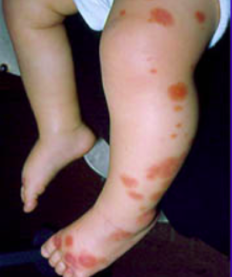
Figure 1. “Henoch-Schonlein Purpura.” Healthychildren.org, American Academy of Pediatrics, 9 Mar. 2017, www.healthychildren.org/English/health-issues/conditions/skin/Pages/Henoch-Schonlein-Purpura.aspx.
CBC, BMP, coags, and UA were obtained. Blood cultures were deferred as patient was not febrile or toxic on presentation. Labs were all within normal limits.
DIAGNOSIS
IgA Vasculitis (formerly known as “Henoch-Schönlein Purpura”)
DISCUSSION
Differentiating Rashes
Skin lesions in children can be daunting. Recognizing if a lesion is blanching or non-blanching can help differentiate rashes. Non-blanching lesions don’t change color with pressure because bleeding is within the skin, not within vasculature. These types of lesions can be distinguished based on size. Petechiae are small (1-3mm), pinpoint non-blanching lesions whereas, purpura are larger (3-5mm), non-blanching lesions and are often raised. The differential for non-blanching lesions is broad. In emergency medicine, it is important to first consider can’t miss diagnoses such as, meningococcus, DIC, malignancy and non-accidental trauma, as these are life threatening. Alternatively, differential considerations can be thought based on etiology, such as: Infectious (meningococcal, endocarditis, RMSF), traumatic (accidental, NAT, increased pressure from coughing, vomiting, straining), hematologic (leukemia, ITP, DIC, HUS), Vasculitis (HSP). There are other causes that are rare and often not diagnosed in the emergent setting which we will not discuss. To differentiate between these etiologies, lab work is often obtained. CBC can be used to look for the presence or absence of thrombocytopenia or signs of infection or large blood cell abnormalities that might point to leukemia or malignancy. PT/PTT/INR can help look for alterations in clotting cascade. BMP/LFTs can be useful if concerned about DIC, HSP, HUS. Blood cultures should be obtained in febrile or toxic patients [5].
What is IgA Vasculitis?
IgA vasculitis is a systemic autoimmune vasculitis where antibodies (IgA) deposit within small blood vessels leading to inflammation and subsequent damage in the form of leakage and bleeding [1,2,3,4]. Technically since it is a systemic disease it can affect all small blood vessels but, IgA vasculitis has a propensity to deposit in blood vessels within the skin, GI tract, joints and kidneys. These deposits lead to the common clinical findings associated with IgA vasculitis including rash, abdominal pain, arthalgias and kidney disease.
Clinical manifestations:
Rash is often the first clinical manifestation of IgA vasculitis [1]. When skin vessels are damaged by IgA deposits, blood leaks into the dermis causing the characteristic rash of IgA vasculitis. The rash is described as a non-blanching, palpable, erythematous rash often involving gravity dependent regions such as the lower extremities and buttock [1, 2, 3, 4]. It typically appears in crops and is occasionally itchy but not often painful [1]. The rash lasts approx. 1-2 weeks.

Figure 2. “IgA Vasculitis Skin Manifestation Examples.” Henoch-Schonlein Purpura, KidsHealth NZ, 2020, www.kidshealth.org.nz/henoch-schonlein-purpura.
Arthralgias (painful, swollen joints) occur in approximately 80% of patients but are often not the presenting symptom. The arthralgias are usually transient or migratory often affecting large lower extremity joints such as the hips, knees and ankles. The upper extremities can be affected as well but are less common. Joint pain may present in younger children as refusal to ambulate. The arthralgias are temporary and do not lead to chronic damage [1].
GI symptoms occur in approx. 50% of patients [1]. The symptoms are caused by inflammation and edema within the GI tract and range from common symptoms such as nausea, vomiting, diarrhea/constipation to severe symptoms such as bowel obstruction, intussusception and GI bleeding [3].
Kidney involvement is often seen through testing of kidney function and UA rather than through reported symptoms. Approximately 25-50% of children develop renal involvement [1]. The kidneys are affected through IgA deposits in the glomerulus capillaries affecting the filtration of the kidney. This damage leads to altered filtration allowing more RBC and protein to exit the body through the urine. This manifests as hematuria and/or mild proteinuria [2]. Rarely, kidney damage will be manifested as high blood pressure and facial swelling.
What causes IgA vasculitis?
IgA vasculitis is classified as an autoimmune vasculitis, meaning the body’s natural defense mistakenly attacks healthy tissue [2]. This occurs when IgA is deposited and the body’s immune system sends in cells to fight off the “intruder”, which leads to inflammation and damage as previously mentioned. The trigger that causes the deposition in the first place is largely unknown. Possible triggers include medications, immunizations, and environmental factors such as food and insect bites [1].
Epidemiology: Who gets IgA vasculitis?
IgA vasculitis is the most common systemic vasculitis in children, with 90% of patients falling within the pediatric age group. The average range differs amongst sources but is around 3-17 years old. The annual incidence is 10-20/100,000 children [1].
How do you diagnose IgA vasculitis?
IgA vasculitis is mainly a clinical diagnosis. There is no one blood test to specify if it is indeed IgA vasculitis. There are many blood tests that point to the diagnosis, such as normal platelet count and coagulation studies, and possibly elevated BUN/Cr and UA with proteinuria/hematuria in a patient with the above-mentioned clinical picture. However, to be fully sure of the diagnosis, a biopsy is needed which would show IgA deposits [4]. This is often unnecessary, as IgA vasculitis is a self-limiting disease with rare long lasting repercussions.
What are the treatment options?
Most people recover from IgA vasculitis without treatment in weeks to months depending on the patient [3]. If treatment is needed it is mainly guided towards symptom management such as NSAIDs for joint pain. If abdominal symptoms are prevalent, a course of corticosteroids such as prednisone can be considered to reduce inflammation in the GI tract, however, the clinician must consider risk vs. benefit in medications with high side effect profile [1]. When serious kidney involvement occurs, patients may need a short course of immunosuppressants.
Complications of IgA vasculitis:
Like previously stated, this is usually a self-resolving disease process. Serious complications include intussusception and bowel obstruction when the inflammation in the GI tract acts as a lead point for telescoping of the bowel. In patients with severe abdominal pain with the classic pruritic rash, intussusception must be considered and an ultrasound should be performed. Rarely does kidney involvement lead to CKD [1]. If a patient has persistent hypertension, proteinuria and/or swelling, a thorough evaluation of IgA nephropathy should be pursued and involvement of a pediatric nephrologist considered.
CASE RESOLUTION
The patient had no lab abnormalities and no signs of renal involvement or symptoms of bowel obstruction. An US of the abdomen was obtained to ensure no evidence of intussusception given the intermittent abdominal pain. That was normal. The patient did not require any treatment in the ED. He was referred to his PCP to monitor symptoms. Mom was instructed to give NSAIDs/acetaminophen for any discomfort and counseled on signs/symptoms of intussusception as reasons to return to the emergency department.
TAKE-AWAYS
If you see non-blanching lesions in children, always consider the can’t miss diagnoses: meningococcus, DIC, HUS, NAT, and leukemia
IgA vasculitis is the most common vasculitis in pediatrics
The most commonly affected regions include: skin, joints, GI tract and kidneys
Think about intussusception and obstruction in children presenting with purpuric rash and abdominal pain
IgA vasculitis is usually self-resolving and treatment typically involves symptom management
AUTHOR: Dr. Kathleen Miller is a current second year emergency medicine resident at Brown University/Rhode Island Hospital.
FACULTY REVIEWER: Dr. Meghan R. Beucher is assistant professor of emergency medicine and pediatrics and Brown University/Hasbro Children’s Hospital.
CITATIONS
Dedeoglu, Fatima, and Susan Kim. “IgA Vasculitis (Henoch-Schönlein Purpura): Clinical Manifestations and Diagnosis.” UpToDate, 2019, www.uptodate.com/contents/iga-vasculitis-henoch-schonlein-purpura-clinical-manifestations-and-diagnosis.
“IgA Vasculitis (Formerly Henoch-Schönlein Purpura or HSP).” UNC Kidney Center, UNC School of Medicine, 6 Apr. 2018, unckidneycenter.org/kidneyhealthlibrary/glomerular-disease/iga-vasculitis-formerly-henoch-schonlein-purpura-or-hsp/.
“IgA Vasculitis.” National Institute of Diabetes and Digestive and Kidney Diseases, U.S. Department of Health and Human Services, 1 Apr. 2020, www.niddk.nih.gov/health-information/kidney-disease/iga-vasculitis.
“IgA Vasculitis.” Vasculitis Foundation, Vasculitis Foundation, 1 Apr. 2019, www.vasculitisfoundation.org/education/forms/henoch-schonlein-purpura/.
McGrath, Ailbhe. “Petechiae.” StatPearls [Internet]., U.S. National Library of Medicine, 10 Aug. 2020, www.ncbi.nlm.nih.gov/books/NBK482331/.