Red Hot in the Age of COVID: Comparing MIS-C to Kawasaki Disease
CASE
A 6-month-old vaccinated, previously healthy boy presented to the ED with fever, rash, and irritability. He had fever for four days (Tmax 102°F). Three days prior, he also developed a red non-pruritic rash and oral lesions. The rash started on his face and progressed to the rest of his body, sparing his palms and soles. His eyes have been red and irritated. He had decreased oral intake and poor urine output. His mother had been giving acetaminophen and ibuprofen. There had been no known sick or COVID-19 contacts.
Vital signs in the ED were notable for blood pressure of 96/79, heart rate of 200, temporal temperature of 38.1 °C (100.6 °F), respiratory rate of 70, and oxygen saturation of 99%.
Physical exam was notable for an irritable but active boy. He has conjunctival injection, dry mucous membranes, erythematous tongue with prominent papillae, and cracked erythematous lips. No lymphadenopathy was present. Heart and breath sounds were normal. Skin exam was notable for diffuse blanching erythematous coalescing patches on face, trunk, extremities and GU area sparing palms and soles. Hands and feet were swollen. Capillary refill was less than 2 seconds.
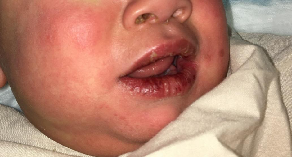
Figure 1. Cracked lips and erythematous rash on face.

Figure 2. Diffuse rash with erythematous patches.
Laboratory evaluation was notable for elevated CRP (136.9 mg/L) and elevated D-dimer (724 ng/mL). Compete metabolic panel was notable only for mildly elevated ALT (76 IU/L). CBC was normal. Troponin was normal and BNP was mildly elevated at 55.8 pg/mL. Urinalysis with 31 WBC’s, trace leukocyte esterase, no nitrites (catharized specimen). Blood and urine cultures and a respiratory pathogen panel (includes COVID-19 testing) were sent.
DIAGNOSIS
This child presented with four days of a febrile illness. His exam was notable for tachycardia, rash, conjunctivitis. The differential diagnosis for this patient was broad and included: Kawasaki disease, multisystem inflammatory syndrome in children (MIS-C), staphylococcal or streptococcal toxic shock syndrome, bacteremia, viral infection, pneumonia, UTI. The patient continued to have fevers after admission and was diagnosed with Kawasaki disease.
DISCUSSION
Kawasaki Disease
Kawasaki Disease (KD) is an acute, self-limited vasculitis that can lead to coronary artery aneurysm and cardiovascular shock. It is the most common cause of acquired heart disease in developed countries. KD is typically seen in children younger than 5 years old, most commonly in children of Asian descent and least commonly in Caucasian children. [1] It is postulated that an infectious agent enters the respiratory tract and triggers an inflammatory cascade. [2] This hyper-inflammatory state affects cardiac tissue leading to myocarditis, arteritis, and coronary aneurysm, resulting in reduced ejection fraction and cardiogenic shock. [1]
Children typically present with multiple days of fever and malaise, which can last 1-2 weeks. Cardiac involvement usually occurs 4-6 weeks after initial symptoms. [1]
Criteria for KD published by the American Heart Association (AHA) are as follows [2]:
Fever for 5 days or more.
At least 4 of the following criteria:
Bilateral painless, nonexudative conjunctival injection, with limbic sparing (figure 3).
Erythematous mouth, strawberry tongue or red, cracked lips
Polymorphous exanthem
Swelling of hands and feet with erythema of the palms and soles
Cervical lymphadenopathy

Figure 3. Conjunctival injection with limbic sparing.
Atypical Kawasaki Disease
The criteria for KD above does not identify all affected children. The diagnosis of atypical KD should be considered in children with prolonged unexplained fever, less than 4 of the AHA criteria, compatible laboratory findings, or concerning echocardiographic findings (such as coronary artery aneurysm or reduced EF) (Figure 4). [2]
![Figure 4. Proposed algorithm for the evaluation of suspected incomplete Kawasaki disease [2].](https://images.squarespace-cdn.com/content/v1/56e8a86a746fb97ea9d14740/1618452052751-5FAY31PFSRYE70QCYJLB/chart+kawasaki.png)
Figure 4. Proposed algorithm for the evaluation of suspected incomplete Kawasaki disease [2].
Treatment
Treatment is targeted at suppressing the inflammatory response and preventing cardiac involvement. Patients should receive high dose IVIG as well as high dose aspirin until the patient becomes afebrile. [1] Corticosteroids may decrease risk of developing cardiac abnormalities and duration of hospitalization. Most studies have shown no harm to adding steroids. [1, 3, 4]
Treatment of KD in the emergency department should be targeted at stabilization with intravenous fluids, vasopressors, and antipyretics. While inpatient, cardiology and infectious disease consultation is recommended.
MISC-C
Presentation/Pathophysiology
Since April 2020, several cases of hyperinflammatory shock linked to COVID-19 in children have been reported. This syndrome has many overlapping features with KD, but is defined as Multisystem Inflammatory Syndrome in Children (MIS-C). [5] As the name suggests, it is characterized by multi-organ involvement which typically includes fever, rash, conjunctivitis, gastrointestinal symptoms, and elevated markers of inflammation. MIS-C is a hyperinflammatory shock state that occurs typically 2-4 weeks after COVID-19 infection. [5] Though many of the symptoms between COVID-19 MIS-C and KD overlap, age of the patient and the presence of COVID-19 antibodies are distinguishing features. MIS-C is usually seen in older children and adolescents, whereas KD is often seen in children under 5 years. [6] An increased prevalence of MIS-C in children of African and Hispanic descent was noted in several studies. [5,6]
The CDC provides the following definition for MISC-C [7]:
Patient aged <21 years
Fever >24 hours
Laboratory evidence of inflammation (elevated CRP, ESR, fibrinogen, procalcitonin, d-dimer, ferritin, LDH, or IL-6, elevated neutrophils, reduced lymphocytes and low albumin)
evidence severe illness requiring hospitalization with multisystem (>2) organ involvement (cardiac, renal, respiratory, hematologic, gastrointestinal, dermatologic or neurological)
AND
No alternative plausible diagnosis
AND
Positive for recent COVID-19 infection or COVID-19 exposure within four weeks of symptom onset.
There is a wide array in the severity of illness for MIS-C, ranging from clinically stable to decompensated shock. In one study of 33 cases, reduced ejection fraction was seen in 63% of patients. [6] It is thought that MIS-C is an antibody-mediated cytokine storm that results in myocardial injury, including coronary aneurysms, similar to KD [8, 9, 10].
Treatment
Though there is much still to learn about MIS-C, elevated inflammatory markers suggest a role of immunomodulatory therapies. In several studies, IVIG and corticosteroids were given to patients. [6, 12] Tocilizumab, a humanized monoclonal antibody against the IL-1 and IL-6 receptors, was also used as these patients often had high levels of these cytokines. [6, 11, 12] IVIG and Tocilizumab may take time to be prepared and may not have immediate effect. As such, the emergency physician’s priority is to stabilize the patient. Patients may require mechanical ventilation, pressor support, and even ECMO in some cases. [6] All emergency physicians typically have access to steroids, and there should be strong consideration for administration in concerning cases. With supportive and immunomodulatory care, most patients do well with normalization of cardiac function. [5]
CASE RESOLUTION
The patient’s presentation, specifically four days of fever, conjunctival injection, cracked lips, and rash raised concern for the diagnosis of KD. Given the COVID-19 pandemic, MIS-C was also in the differential. Given initial tachycardia, bedside echocardiogram was performed, which was normal. IV fluids and antipyretics were administered in the ED and the patient’s hemodynamics improved. While admitted to the floor, he continued to have up to 5 days of fever, fulfilling the criteria for KD. He was started on IVIG, high dose ASA, and steroids. COVID-19 antibody testing was negative. After 4 days of treatment and monitoring, he was discharged in good condition with aspirin and a steroid taper.
AUTHOR: Camila Tyminski, MD is a second year emergency medicine resident at Brown University/Rhode Island Hospital.
FACULTY REVIEWERS: Meghan Beucher, MD and Alicia E. Genisca, MD are pediatric emergency medicine physicians at Hasbro Children’s Hospital
REFERENCES
Modesti AM, Plewa MC. Kawasaki Disease. [Updated 2020 Jul 2]. In: StatPearls [Internet]. Treasure Island (FL): StatPearls Publishing; 2020 Jan-.
McCrindle BW, Rowley AH, Newburger JW, Burns JC, Bolger AF, Gewitz M, Baker AL, Jackson MA, Takahashi M, Shah PB, Kobayashi T, Wu MH, Saji TT, Pahl E., American Heart Association Rheumatic Fever, Endocarditis, and Kawasaki Disease Committee of the Council on Cardiovascular Disease in the Young; Council on Cardiovascular and Stroke Nursing; Council on Cardiovascular Surgery and Anesthesia; and Council on Epidemiology and Prevention. Diagnosis, Treatment, and Long-Term Management of Kawasaki Disease: A Scientific Statement for Health Professionals From the American Heart Association. Circulation. 2017 Apr 25;135(17):e927-e999.
Saguil A, Fargo M, Grogan S. Diagnosis and management of Kawasaki disease. Am Fam Physician. 2015 Mar 15;91(6):365-71.
Miyata K, Kaneko T, Morikawa Y, Sakakibara H, Matsushima T, Misawa M, Takahashi T, Nakazawa M, Tamame T, Tsuchihashi T, Yamashita Y, Obonai T, Chiga M, Hori N, Komiyama O, Yamagishi H, Miura M., Post RAISE group. Efficacy and safety of intravenous immunoglobulin plus prednisolone therapy in patients with Kawasaki disease (Post RAISE): a multicentre, prospective cohort study. Lancet Child Adolesc Health. 2018 Dec;2(12):855-862.
Riphagen S, Gomez X, Gonzalez-Martinez C, Wilkinson N, Theocharis P. Hyperinflammatory shock in children during COVID-19 pandemic. Lancet 2020;395:1607–8. 10.1016/S0140-6736(20)31094-1
Kaushik S, Aydin SI, Derespina KR, et al. Multisystem Inflammatory Syndrome in Children Associated with Severe Acute Respiratory Syndrome Coronavirus 2 Infection (MIS-C): A Multi-institutional Study from New York City. J Pediatr. 2020;224:24-29. doi:10.1016/j.jpeds.2020.06.045
Centers for Disease Control and Prevention Emergency preparedness and response. https://emergency.cdc.gov/han/2020/han00432.asp
Fujimaru T., Ito S., Masuda H., Oana S., Kamei K., Ishiguro A., Kato H., Abe J. Decreased levels of inflammatory cytokines in immunoglobulin-resistant Kawasaki disease after plasma exchange. Cytokine. 2014;70:156–160. doi: 10.1016/j.cyto.2014.07.003
Tunuguntla H., Jeewa A., Denfield S.W. Acute myocarditis and pericarditis in children. Pediatr Rev. 2019;40:14–25
Toubiana J, Poirault C, Corsia A, et al. Kawasaki-like multisystem inflammatory syndrome in children during the covid-19 pandemic in Paris, France: prospective observational study. BMJ. 2020;369:m2094.
Zhang C., Wu Z., Li J.W., Zhao H., Wang G.Q. The cytokine release syndrome (CRS) of severe COVID-19 and interleukin-6 receptor (IL-6R) antagonist tocilizumab may be the key to reduce the mortality. Int J Antimicrob Agents. 2020;55:105954.
Cheung EW, Zachariah P, Gorelik M, et al. Multisystem Inflammatory Syndrome Related to COVID-19 in Previously Healthy Children and Adolescents in New York City. JAMA. 2020;e2010374.