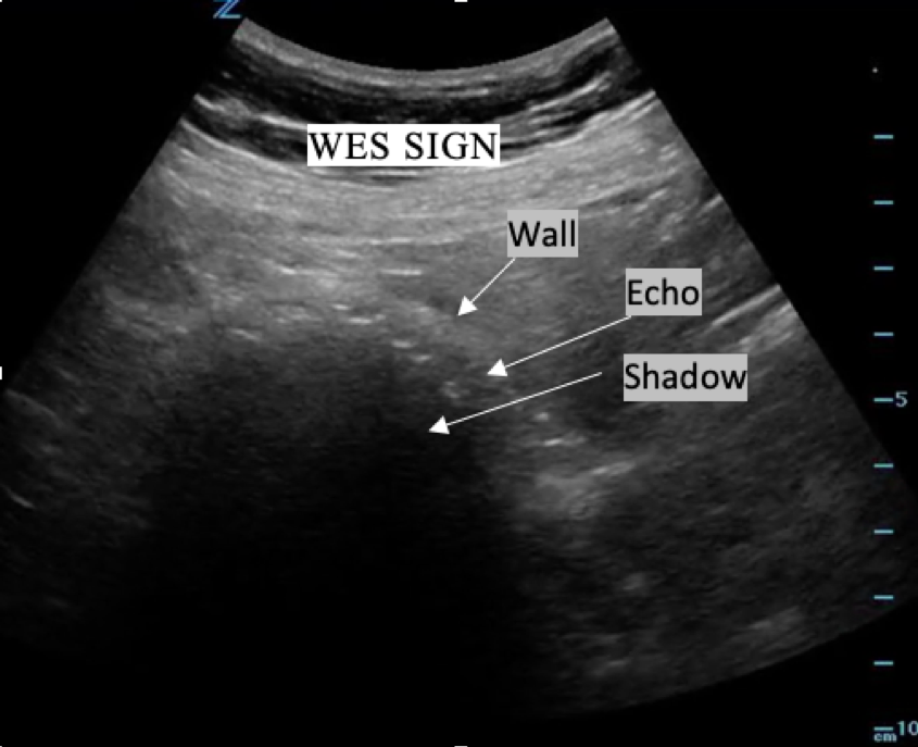
Avoiding a Pitfall of Biliary Ultrasound: Understanding Wall-Echo-Shadow (WES) Sign
CASE:
40 year old female with normal vital signs and a chief complaint of right upper quadrant abdominal pain associated with nausea and vomiting. She has never had this pain before. She has no past medical and no past surgical history. She denies vaginal and urinary symptoms. She undergoes right upper quadrant ultrasound which confirms the diagnosis.
DIAGNOSIS:
Cholelithiasis.
DISCUSSION:
While the differential diagnosis of abdominal pain is broad, in this particular case, cholelithiasis and cholecystitis are likely near the top of the list of pathologies being considered. For this reason, bringing the ultrasound machine along as you begin your initial evaluation is prudent.
The curvilinear probe is the probe of choice for a biliary ultrasound. In regards to technique, ACEP’s Sonoguide instructs, “Place the probe in sagittal (longitudinal) orientation with the probe-indicator oriented toward the patient’s head and instruct the patient to take a deep breath. Sweep the probe inferiorly and laterally along the right subcostal margin” [1]. On this patient, that technique does not allow for visualization of the gallbladder, and so a second approach is used. The probe is placed in the longitudinal orientation on the patient’s right lateral body, with probe-indicator toward the patient’s head, creating a coronal view. This is the same view used in the FAST exam. Once the kidney is visualized, fanning the probe anteriorly should allow the gallbladder to come into view. However, on this patient, this technique also fails. On the screen, instead of a gallbladder, there is a hyperechoic line followed by a large shadow. To the unknowing eye, one may assume this is bowel; and thus, the shadowing being caused by bowel gas. However, if the correct techniques to identify the gallbladder have been used, and landmarks such as the portal triad have been identified, Wall-Echo-Shadow (WES) sign may be present. The differential for difficulty visualizing the gallbladder with ultrasound includes cholecystectomy, fasting, recent meal, and WES sign.
More specifically, the WES sign is “…the demonstration of the gallbladder Wall, the Echo of the stone, and the acoustic shadow” [2]. This triad should be used to “differentiate the contracted gallbladder with stones from a loop of bowel containing gas”[2]. On your ultrasound screen, you will see “…two curvilinear, parallel echogenic lines separated by a thin hypoechoic space and acoustic shadowing distal to the echogenic line in the far field” beneath the gallbladder wall, and prominent posterior acoustic shadowing that results from sound attenuation caused by the calculi. The hypoechoic region between the echogenic gallbladder wall and subjacent calculi represents a thin layer of interpositioned bile” [3].
CASE RESOLUTION:
This patient received a general surgery consult and was admitted for a cholecystectomy.
TAKE-AWAYS:
Comfort in recognizing WES sign is imperative prior to performing biliary ultrasounds in the clinical setting. In understanding that the sensitivity of bedside ultrasound by emergency physicians is over 85% [1] if WES sign were mistaken for bowel, one might conclude gallstones to be unlikely cause of a patient’s pain. This would lead to delay in diagnosis and delay to definitive care. With that understanding, my approach to a difficult biliary ultrasound starts with first asking the patient if they are sure that they still have their gallbladder, inquiring about recent meals, and lastly, looking carefully for WES sign.
Keywords: WES Sign, gallbladder, biliary, cholelithiasis, cholecystitis
Author: Kaitlin Lipner, MD is a first year resident at Brown University/Rhode Island Hospital
Faculty Reviewer: Kristin Dwyer, MD is the Director of Ultrasound Division of Brown Emergency Medicine
References:
Sonoguide [Internet]. American College of Emergency Physicians. Gallbladder. 2020 [accessed 2021 June 5]. Available from: https://www.acep.org/sonoguide/basic/gallbladder/
MacDonald, F. R., et al. “The WES Triad — A Specific Sonographic Sign of Gallstones in the Contracted Gallbladder.” Gastrointestinal Radiology, vol. 6, no. 1, 1981, pp. 39–41., doi:10.1007/bf01890219.
Rybicki, Frank J. “The WES Sign.” Radiology, vol. 214, no. 3, Mar. 2000, pp. 881–882., doi:10.1148/radiology.214.3.r00mr38881.
