Red, Hot, Legs: Thrombophlebitis of the Superficial Veins of the Lower Extremity
CASE
A 34-year-old female with medical history notable for varicose veins presents with complaint of left medial thigh pain. The patient states the pain started 3 days ago. She denies any fevers. She states the area is very painful to the touch. No antecedent trauma or injury. She has been taking Tylenol with minimal relief. She has a remote history of varicose vein ablation to her left leg.
Vital Signs: HR 85 BP 120/80 T 99F RR 20
Physical Exam
Left Lower Extremity: Warm, erythematous, indurated area measuring 7 cm along left medial thigh. No fluctuance. No crepitus. No vesicular lesions. No blisters/bullae. Scattered varicose veins in bilateral lower extremities. Normal distal pulses.
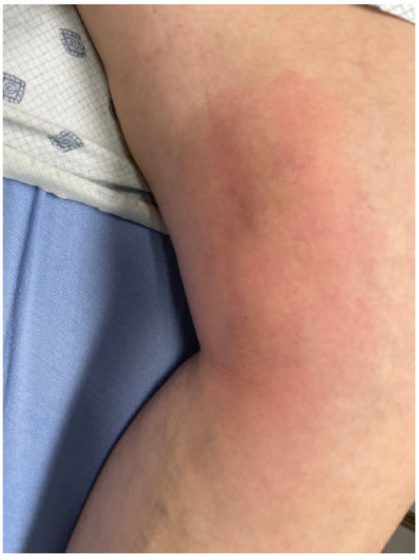
Image 1. LLE Exam Findings
Differential Diagnosis
Cellulitis/Erysipelas, abscess, contact/allergic dermatitis, superficial phlebitis, superficial thrombophlebitis , superficial vein thrombosis (SVT)
ED Evaluation
An ultrasound was performed which showed no evidence of deep vein thrombosis involving the left lower extremity and thrombosed varicosities of the medial left thigh, consistent with superficial thrombophlebitis, involving the greater saphenous vein.
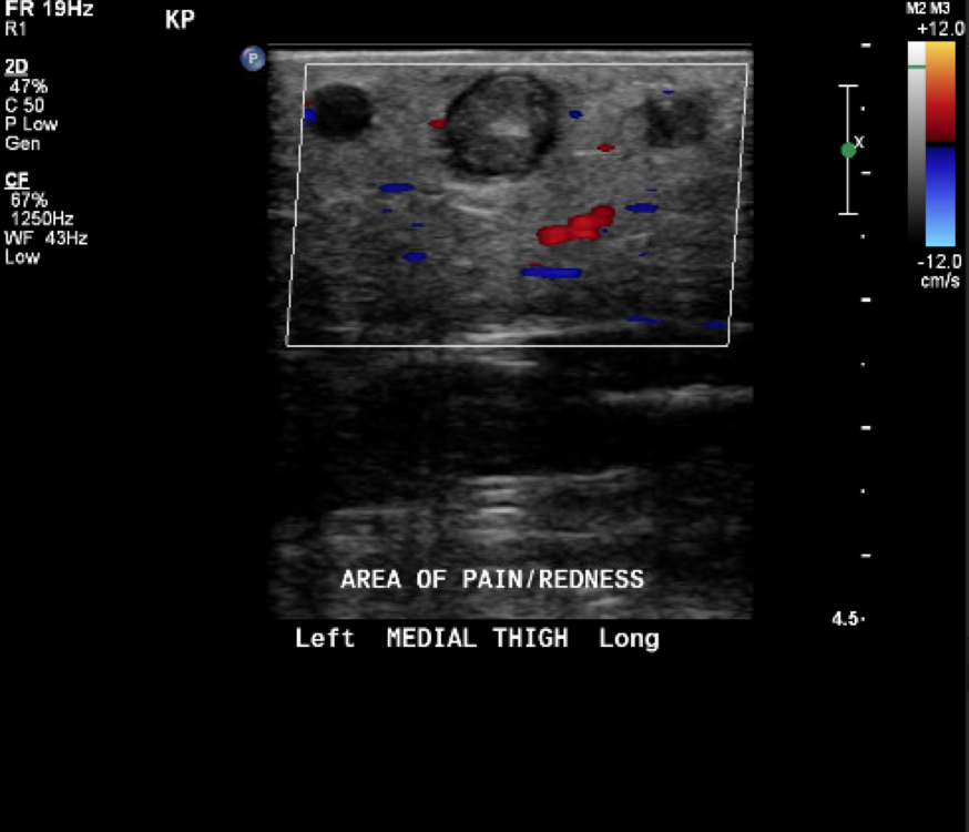
Image 2. Ultrasound findings
DIAGNOSIS
Superficial Vein Thrombosis (SVT) of the greater saphenous vein.
DISCUSSION
First, let’s discuss definitions.
Superficial phlebitis describes pain and inflammation within a vein without any associated thrombus. Superficial thrombophlebitis describes inflammation and a thrombus within the tributary veins of the lower extremity. Superficial vein thrombosis (SVT) describes inflammation and thrombus within the axial veins of the lower extremity. The axial veins of the lower extremity are the great saphenous, accessory saphenous, and small saphenous.

Image 3. Anatomy of superficial veins of lower extremity
One more condition to familiarize yourself with is suppurative thrombophlebitis. This describes venous inflammation and thrombosis within an infected vein. This condition is associated with fever, fluctuance and purulence. This condition rarely occurs without a preceding history of cannulation/catherization of the affected vein. This condition necessitates antibiotics.
Although there is obvious clinical overlap in these conditions, the differentiation is of great importance. SVT is associated with increased risk for propagation into the deep venous system and heightened potential for development of pulmonary embolism. Patients with SVT were found to have an 8.3% risk of complications at 3 months including PE (0.5%), DVT (2.8%), extension of SVT (3.3%), and recurrence of SVT (1.9%) [1].
Recognition and initiation of the appropriate treatment for these various inflammatory/thrombotic conditions is of paramount importance to our specialty. Treatment options include conservative management (NSAIDs, compression stockings), anticoagulation for 45-days (prophylactic anticoagulation) or anticoagulation for durations consistent with those used in the management of DVT (termed therapeutic anticoagulation).
Treatment must be individualized and focuses on many factors including patient risk factors, location of the clot and size of the clot.
Let’s break this down.
What is the patient risk?
Patient with increased risk for VTE should be considered high-risk. This includes patients with history of malignancy, estrogen use, immobility, etc.
Patients are also considered high-risk if they have had propagation of SVT despite anticoagulation or recurrence of SVT after discontinuation of anticoagulation.
Patients are considered low-risk if they have no medical risk factors for VTE and demonstrate appropriate therapeutic response with conservative management.
What is the location risk?
Axial veins are higher risk than tributary veins (the difference between superficial thrombophlebitis and superficial vein thrombosis)
SVT within the great saphenous, accessory saphenous, and small saphenous that is within 3 cm of the saphenofemoral (SFJ) or saphenopopliteal (SPJ) junction is considered high risk
SVT is considered intermediate risk if located within 3-5 cm of the deep venous system (i.e. located within 3-5 cm of the SFJ or SPJ)
What is the size risk?
A clot involving a >5 cm portion of a vein is considered intermediate-risk.

To summarize, low risk patients can be managed with NSAIDs (as tolerated), compression stockings and repeat US (guidelines recommend within 2-3 days if managing SVT and within 7-10 days if following superficial thrombophlebitis/phlebitis). Intermediate risk patients should be treated with 45-days of prophylactic anticoagulation. High-risk patients should be treated with therapeutic anticoagulation (same duration as if treating for a DVT) [3].
The choice of anticoagulation is debated. The CALISTO trial, from which much of the current management guidelines is extrapolated, used fondaparinux as their anticoagulant of choice. The SURPRISE trial showed noninferiority of 10 mg of rivaroxaban (prophylactic dose) compared with fondaparinux. Cost, ease of use and patient preference should be factored into anticoagulant choice [2].
CASE RESOLUTION
Our patient was considered intermediate risk. Our patient was started on 45 days of anticoagulation with Eliquis.
Radiology does not always document the size or location (as in distance from SPJ, SFJ) of SVT. Given the clinical implications, please consider contacting the radiologist to appropriately risk stratify patients.
TAKE-AWAYS
The differential for a red, warm leg is broad. Understanding the difference between superficial phlebitis, superficial thrombophlebitis and superficial vein thrombosis has implications on treatment.
Based on ultrasound and patient history, place patient into low, intermediate or high-risk categories.
Anticoagulation for 45 days is recommended for intermediate risk patients. Therapeutic anticoagulation is recommended for patient’s at high risk.
Keywords:
Superficial vein thrombosis, superficial thrombophlebitis
AUTHOR: Michael Tcheyan, MD is a third-year emergency medicine resident at Brown University.
FACULTY REVIEWER: Madelene Boyle, MD is an attending emergency medicine physician at Newport Hospital.
REFERENCES
Decousus H, Quéré I, Presles E, Becker F, Barrellier MT, Chanut M, Gillet JL, Guenneguez H, Leandri C, Mismetti P, Pichot O, Leizorovicz A; POST (Prospective Observational Superficial Thrombophlebitis) Study Group. Superficial venous thrombosis and venous thromboembolism: a large, prospective epidemiologic study. Ann Intern Med. 2010 Feb 16;152(4):218-24. doi: 10.7326/0003-4819-152-4-201002160-00006. PMID: 20157136.
Werth S, Bauersachs R, Gerlach H, Rabe E, Schellong S, Beyer-Westendorf J. Superficial vein thrombosis treated for 45 days with rivaroxaban versus fondaparinux: rationale and design of the SURPRISE trial. J Thromb Thrombolysis. 2016 Aug;42(2):197-204. doi: 10.1007/s11239-016-1354-3. PMID: 26973347.
Scovell, Sherry, et al. (2021). Superficial Vein Thrombosis and Phlebitis of the Lower Extremity Veins, UpToDate. Retrieved December 11, 2021.