FAST TRACK: Auricular Hematomas
CASE
An 18-year-old male presents from a boxing match with right external ear pain and swelling after being struck in the head. He denies other injuries. Hearing is intact but on exam there is significant pain, swelling and deformity of the pinna.
DIAGNOSIS
Auricular hematoma
DISCUSSION
Auricular hematomas are deformities to the pinna typically caused by blunt trauma, resulting in separation of the perichondrium from underlying cartilage and tearing of adjoining blood vessels. On exam there is loss of typical auricular landmarks and severe pain. Cartilage is dependent on the perichondrium for vascularity, thus auricular hematomas that are not treated promptly may cause cartilage deformities and long-lasting disfigurement of the ear (cauliflower ear) [1].

Figure 1: A) Auricular hematoma [1] and B) cauliflower deformity [2]
Auricular hematomas may be treated with incision and drainage. Needle aspiration may be attempted but is generally insufficient to evacuate larger hematomas where clot has already formed [3]. Indication for drainage is traumatic swelling with pinna deformity on exam, within 7 days of trauma occurring. Contraindications for drainage include recurrent or chronic hematoma, > 7 days from the trauma, or evidence of infection. In these cases, referral to an ENT or a plastic surgeon should be made.
Incision & Drainage:
Equipment Needed:
PPE: sterile drapes, gown, face/eye protection, gloves
5cc syringe
20-gauge needle or bigger
Topical antiseptic/iodine
1% lidocaine
Scalpel (#11 blade)
Hemostat and forceps
Dental rolls
Gauze packing and bandages
Xeroform
Laceration tray setup: needle driver, 3-0 nonabsorbable suture
Procedural Steps:
Get informed consent from the patient
Place patient in supine position with head slightly turned towards the unaffected side
Perform an auricular block

Figure 2: How to perform an auricular block [6]
4. Evacuate the clot
a. Make a small semi-circle incision along the inner aspect of the curvature of the helix or anti-helix
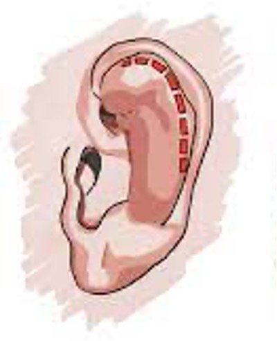
Figure 3 Semicircular incision at the helix that follows the curvature of the ear [7]
b. Evacuate the hematoma by providing pressure from the hematoma towards the incision (“milking” the hematoma).
c. Use a hemostat or needle driver to assist in breaking up hematoma that is difficult to evacuate.
5. Apply an ear bolster to help prevent the auricular hematoma from reaccumulating
a. Consider leaving the incision open to allow for continuous drainage
b. Pack the helix with xeroform
c. Apply dental rolls (or rolled gauze) parallel to the incision at the anterior and posterior aspects of the ear and use a 3-0 suture in place in mattress fashion (see Figure 4).
d. Apply bacitracin ointment to the incision site post-procedure

Figure 4: Ear bolster with dental rolls and mattress suture technique [4]
Follow-up:
Bolster dressing can be removed in 5-7 days and patients should be provided with ENT or plastic surgery follow-up at discharge. Antibiotic use is left to the discretion of the physician, however in immunosuppressed patients, pseudomonal coverage is indicated. Patients should be counseled that reaccumulation of the hematoma is possible, and to limit physical activity and avoid contact sports for 1-2 weeks [5].
TAKE-AWAYS
Auricular hematomas are typically caused by blunt trauma to the pinna
They require prompt treatment to help prevent cartilage deformity and cauliflower ear
Treat with incision and drainage, and apply bolster/splint
Discharge with ENT/plastics follow-up, advise limited activity and caution patients regarding reaccumulation of the hematoma.
Author: Lindsey Brown, MD is a current second-year resident at Brown Emergency Medicine
Faculty Reviewer: Michelle Myles, MD is an attending physician at Rhode Island Hospital and Miriam Hospital.
REFERENCES
1. San, Furkan. “Spontaneous Auricular Hematoma in Behçet Disease.” Entcase, 1 Feb. 2020, entcase.org/manuscript/546/makale/otoloji/spontaneous-aurIcular-hematoma-In-behcet-dIsease.htm. Accessed 13 Feb. 2024.
2. “Cauliflower Ear - Symptoms, Causes and Treatment.” Sportsinjuryclinic.Net, 27 Oct. 2021, www.sportsinjuryclinic.net/sport-injuries/head-face/ear-pain-injuries/cauliflower-ear. Accessed 13 Feb. 2024.
3. Giles WC, Iverson KC, King JD, Hill FC, Woody EA, Bouknight AL. Incision and drainage followed by mattress suture repair of auricular hematoma. Laryngoscope. 2007 Dec;117(12):2097-9.
4. Desai, Bobby K. “Treatment of Auricular Hematoma.” SpringerLink, Springer International Publishing, 1 Jan. 1970, link.springer.com/chapter/10.1007/978-3-030-85047-0_65. Accessed 13 Feb. 2024.
5. Krogmann, Ryan J. “Auricular Hematoma.” StatPearls [Internet]., U.S. National Library of Medicine, 31 July 2023, www.ncbi.nlm.nih.gov/books/NBK531499/. Accessed 13 Feb. 2024.
6. Dinces, Elizabeth A. “How to Do an Auricular Block - Ear, Nose, and Throat Disorders.” Merck Manuals Professional Edition, Merck Manuals, 29 Jan. 2024, www.merckmanuals.com/professional/ear,-nose,-and-throat-disorders/how-to-do-ear-procedures/how-to-do-an-auricular-block. Accessed 20 Feb. 2024.
7. Newman, Thomas. “Ear Trauma.” TeachMeSurgery, 23 Jan. 2024, teachmesurgery.com/ent/ear/ear-trauma/. Accessed 20 Feb. 2024.