Ortho Review: Keepin’ up with the Joneses
Case:
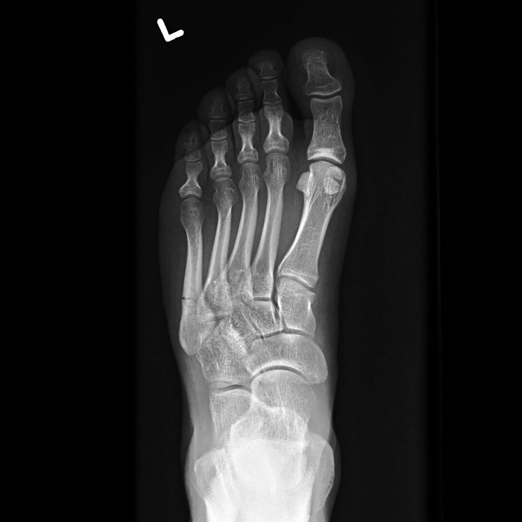
A 31-year-old competitive tennis player presents to the Emergency Department with right foot pain. He was pivoting to chase down the ball on the court when he suddenly developed severe pain on the lateral aspect of his foot. Exam demonstrates swelling and ecchymosis with tenderness to palpation at the base of the fifth metatarsal. Radiographs are obtained (see figure 1 below). What is the appropriate management of this injury?
How do we classify and manage proximal 5th metatarsal fractures in the ED?
Anatomy:

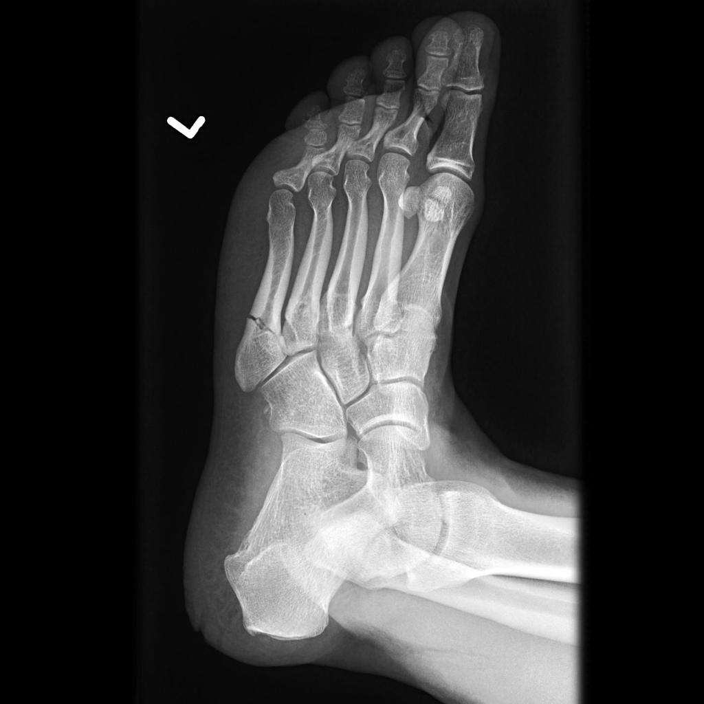
Figure 2: 5th metatarsal anatomy
The three relevant anatomical areas of the 5th metatarsal (proximal to distal) are the proximal tubercle, metaphysis including 4th/5th intermetatarsal facet, and the diaphysis. There are three fracture types, organized into "zones" based upon these three anatomical locations:
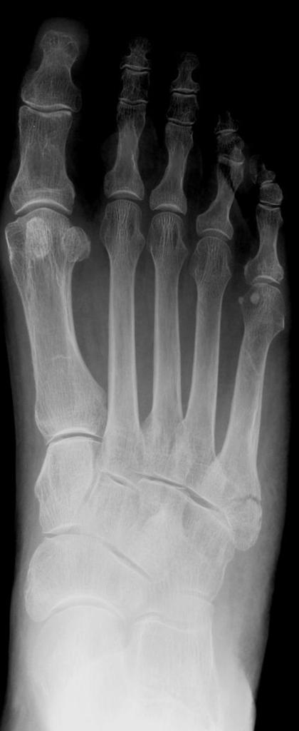
Figure 3: Zone 1 fracture
Zone 1 – Tuberosity avulsion fracture (aka “Pseudo-Jones” or “Dancer’s” fracture)
- Involves the proximal tubercle
- Mechanism: Inversion and plantar flexion (e.g. landing awkwardly after a jump) results in avulsion fracture from the long plantar ligament, lateral band of the plantar fascia, or contraction of the peroneus brevis.
- Blood supply from multiple vessels
- Nonunion is uncommon
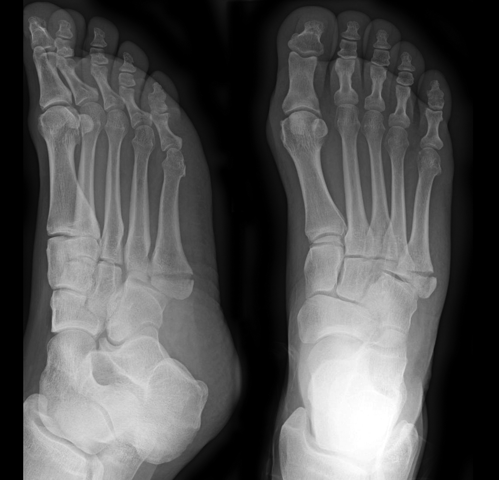
Figure 4: Zone 2 fracture
Zone 2 – The "Jones fracture,” as described by Sir Robert Jones in his 1902 paper, “Fracture of the base of the 5th metatarsal by indirect violence.”
- Occurs at the metaphyseal-diaphyseal junction
- Involves the 4th-5th metatarsal articulation
- Mechanism: Medio-lateral force applied while heel is raised and foot is plantar flexed (e.g. sudden change in direction)
- Vascular watershed area, leading to increased risk of nonunion
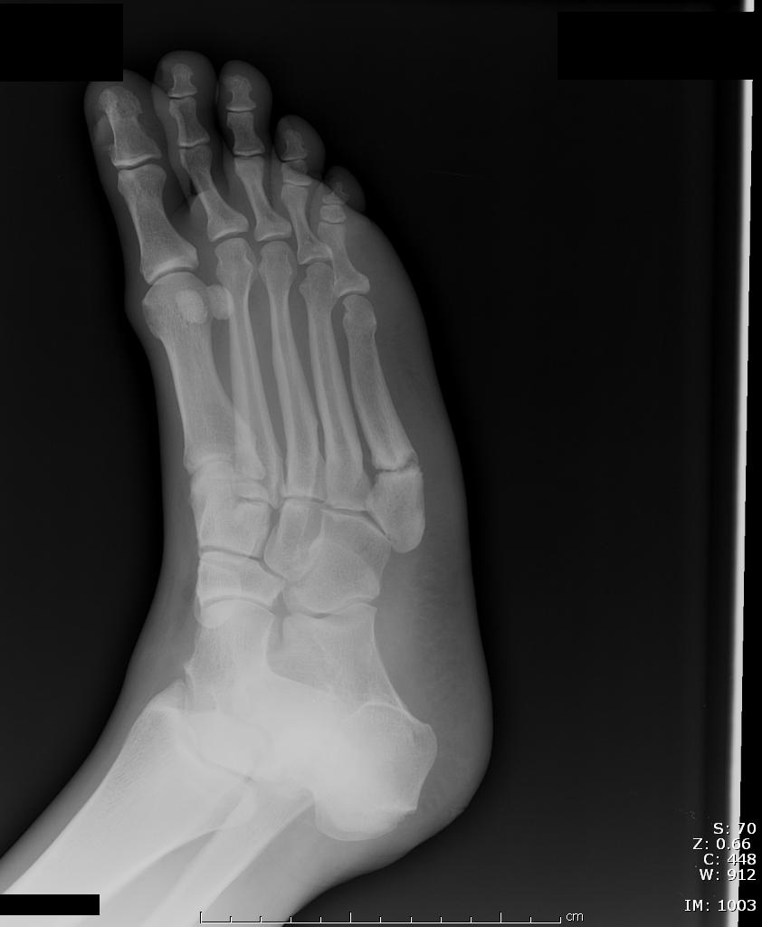
Figure 5: Zone 3 fracture
Zone 3 – Lacking a cool name
- Diaphyseal stress fracture
- Distal to the 4th-5th metatarsal articulation
- Mechanism: Chronic microtrauma, typically involves prodrome of pain for several weeks-months
- As compared to acute, well-defined fracture lines, stress fractures may have widened fracture lines with surrounding cortical thickening
- Associated with cavovarus foot deformities or sensory neuropathies
- Increased risk of nonunion
Presentation and Imaging:
All three injuries present with pain on the lateral edge of the foot, often involving swelling and ecchymosis to the area. These are the fractures we are considering when palpating the base of the 5th metatarsal in applying the extended Ottawa ankle, or Ottawa foot, rules.
Appropriate imaging of the foot includes three views: AP, lateral, and oblique.
Management:
Why should I care? We just toss them in a hard shoe and send them out, right?
Wrong. While we are talking about zones that are millimeters apart, they have vastly different prognoses.
Zone 1 can typically be treated with a hard “post-op” shoe, weight-bearing as tolerated, and Orthopedic follow-up in 1 week.
Zone 2 and zone 3 injuries have an increased risk of nonunion and therefore must be treated with a posterior short leg splint as non-weightbearing injuries. It is of particular importance to ensure prompt orthopedic follow-up for these zone 2 and zone 3 injuries as they will need to be evaluated for possible operative management.
Case Outcome:
Looking closely at the radiographs, the fracture line extends into the intermetatarsal articulation which distinguishes it as a zone 2 injury, or Jones fracture. He was discharged with a splint and instructions for strict non-weightbearing.
Faculty Reviewer: Dr. Jeff Feden
References:
Gaines, SA, Handel, DA, Ramsey, PN. Chapter 274: Foot Injuries. Tintinalli’s Emergency Medicine: A Comprehensive Study Guide. 7th ed. 2011; 1877-1879.
Jones, R. I. Fracture of the Base of the Fifth Metatarsal Bone by Indirect Violence. Ann Surg 1902; 35:697.
Nunley JA. Fractures of the base of the fifth metatarsal: the Jones fracture. Orthop Clin North Am 2001; 32:171.
Mehlhorn AT, Zwingmann J, Hirschmüller A, et al. Radiographic classification for fractures of the fifth metatarsal base. Skeletal Radiol 2014; 43:467.
Weather Ford, Brian. "5th Metatarsal Base Fracture." Orthobullets. Orthobullets.com, 11 Aug. 2015. Web. 31 Mar. 2016.