Case of the Month — Ectopic Pregnancy
CASE
A 26 year-old with no significant past medical history presents with two days of abdominal pain and vaginal bleeding. She describes the pain as a 4/10, “crampy” and diffuse, located in the lower abdomen. She is not experiencing any nausea, vomiting, diarrhea, constipation, dysuria, or increase in urinary frequency or urgency. Her last menstrual period was approximately 7-8 weeks ago and she has a history of regular periods. She has no history of STIs, prior surgeries or pregnancies, and is uncertain whether she desires pregnancy currently.
In the ED, her vitals are normal. Physical exam is notable for mild suprapubic tenderness, no guarding, rigidity, or rebound. Pelvic exam demonstrates a small amount of brown blood in the vault but no clots or active bleeding from her closed cervical os. There is also no obvious uterine enlargement, adnexal tenderness, or cervical motion tenderness on bimanual exam.
Qualitative β-hCG: 8,580 mIU/mL
A transabdominal ultrasound is performed.
DISCUSSION
Vaginal bleeding in pregnancy is a common presenting complaint in the ED, and if this is caused by an ectopic pregnancy, early identification is critical for patient safety. Between the years 2011-2013 for example, ectopic pregnancy accounted for 2.7% of maternal deaths within the US. [1] For patients identified as having an ectopic pregnancy, an initial presentation of 1) positive β-hCG in the first trimester, 2) abdominal pain, and 3) vaginal bleeding were significantly and positively associated with the presence of ectopic pregnancy. [2] Both transabdominal ultrasound (TAUS) and transvaginal ultrasound (TVUS) are helpful in establishing this diagnosis. TVUS in particular has been found to have high sensitivities for the diagnosis of tubal ectopic pregnancy (87.0-99.0%), with specificities ranging from 94.0-99.0%. [3]
TAUS & TVUS Technique
A transabdominal ultrasound (TAUS) should be obtained prior to a transvaginal ultrasound (TVUS), as a transabdominal approach is less invasive for the patient. With TAUS however, appropriate visualization of all structures may be difficult, especially when evaluating for an intrauterine pregnancy (IUP) with earlier gestation. Using the TAUS technique, an IUP is likely to be visualized starting after 6-7 weeks, whereas and IUP may be identified earlier on in gestation using the TVUS approach.

Table 1. Differences between gestational weeks at which TVUS & TAUS can detect signs of early IUP. Obtained from SAEM Lecture Series: Transabdominal Pelvis Ultrasound (Dr. Lorraine Ng)
*These are rough estimates and can be informative, but expect variability. Use cautiously in the clinical context.
For TAUS, a curvilinear or phased array probe should be used and is best performed when the patient has a full bladder to serve as a sonographic window through which more posterior pelvic structures can be viewed. The indicator should be oriented caudally for a sagittal view, then rotated 90 degrees counter-clockwise for a transverse view, with the indicator oriented towards the patient’s right. Pelvic structures should be visualized first, including the bladder, uterus, cervix and vaginal stripe on sagittal view, and Pouch of Douglas, which is the most dependent area in the female pelvis and may be a site of free-fluid accumulation with a ruptured ectopic pregnancy.[4] The adnexa are difficult to assess on TAUS. After identification of these structures, evaluation for the presence of intrauterine contents and/or signs of ectopic pregnancy can be made. The presence of a fetal heart rate can also be measured on M-Mode.
For a TVUS, the bladder should be emptied, and as the majority of patients will have an anteverted uterus, it is helpful to elevate the pelvis if a gynecologic stretcher is not available. The probe of choice for the approach is a sanitized endocavitary probe. Place a small amount of gel on the probe, place a probe cover, then apply a small amount of sterile lubricant onto cover. If the patient prefers to do so, they should be allowed to insert the probe themselves at the start of the exam. Keep the indicator towards the ceiling for a sagittal view — and fan from left to right to view the structures in their entirety. Turn indicator 90 degrees counterclockwise for a coronal view of uterus, fanning up and down. Then rock the probe towards left, and fan up and down, and toward the right adnexa, and fan up and down, to visualize the adnexa to the sides of the uterus.
![Figure 1. Range of locations at which an ectopic pregnancy might be located.[5]](https://images.squarespace-cdn.com/content/v1/56e8a86a746fb97ea9d14740/1614695211282-SCZOL20OCTZJXLRLW5N9/Picture1.png)
Figure 1. Range of locations at which an ectopic pregnancy might be located.[5]
Scenario 1: Intrauterine Pregnancy with Normal Adnexa
The criteria for an intrauterine pregnancy include:
1. Gestational sac
2. Fetal pole or yolk sac
3. IN the uterus and with an appropriate endomyometrial mantle
![Figure 2. First-trimester IUP with gestational sac and yolk sac within the uterine cavity. [6] In this scenario, we see a definitive IUP, and the patient was not on any infertility treatments, making the risk of a heterotopic pregnancy close to zero…](https://images.squarespace-cdn.com/content/v1/56e8a86a746fb97ea9d14740/1614695633858-HV4Q5AP3KVP4ZE3H51GJ/Image+2.png)
Figure 2. First-trimester IUP with gestational sac and yolk sac within the uterine cavity. [6] In this scenario, we see a definitive IUP, and the patient was not on any infertility treatments, making the risk of a heterotopic pregnancy close to zero. Other considerations here include evaluation of the endomyometrial mantle, or the full thickness of the endometrium and myometrium combined. A thickness ≥ 8 mm is considered normal, with < 5 mm concerning for interstitial pregnancy.[7]
![Figure 3. First-trimester IUP with thick endomyometrial mantle. [8]](https://images.squarespace-cdn.com/content/v1/56e8a86a746fb97ea9d14740/1614695791742-BRUTCO199LJ657AGPB8J/Mantle.png)
Figure 3. First-trimester IUP with thick endomyometrial mantle. [8]
Scenario 2: Ectopic Pregnancy Identified, No Intrauterine Pregnancy
If an extrauterine mass is identified containing a gestational sac and a yolk sac and/or fetal pole (with or without cardiac activity), in the absence of an intrauterine pregnancy, the diagnosis of an ectopic pregnancy can be made. In up to 20% of ectopic pregnancies, a pseudogestational sac, or an intrauterine endometrial fluid collection may be visible on ultrasound, and should not be mistaken for a viable IUP.3 A pseudogestational sac structure is characterized by a central location, oval shape, and lack of chorionic ring. The need for timely surgical intervention is guided by clinical presentation. If there is concern for a ruptured ectopic pregnancy, rapid ultrasound detection of free intraperitoneal fluid in Morrison’s pouch has been shown to predict the need for operative intervention. [9] Often, no extrauterine ectopic will be visualized at the bedside, but the absence of a definitive IUP with an elevated hGC and free fluid in Morrison’s pouch as a very high PPV for an ectopic pregnancy requiring surgical intervention.
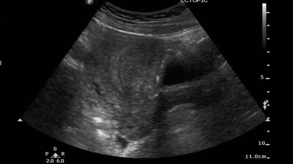
Figure 4. No intrauterine pregnancy visualized.
Scenario 3: Normal Adnexa, No Intrauterine Pregnancy or Findings Concerning for Intrauterine Pregnancy Failure
Here, a relevant concept to consider is the discriminatory zone, or the value at which an intrauterine gestational sac should be visible on ultrasound in a normal pregnancy. More recently, it has been suggested that a single measurement of β-hCG does not reliably distinguish between an ectopic, viable IUP, and nonviable IUP. Rather, in hemodynamically stable patients, trending β-hCG value over 48 hours in conjunction with follow-up imaging is appropriate in order to establish a diagnosis and determine treatment options, if needed. Similarly, guidelines for ultrasound diagnosis below of intrauterine pregnancy failure may be helpful in arriving at a clear diagnosis. [10]

Table 2. Guidelines for TVUS Diagnosis of Intrauterine Pregnancy Failure
TAKE-AWAYS
Without a definitive IUP in a patient with abdominal pain and vaginal bleeding, ectopic pregnancy should be considered with timely diagnosis and treatment being potentially lifesaving.
A gestational sac alone is not sufficient to diagnose an intrauterine pregnancy. Definitive diagnosis of a viable IUP requires identification of a gestational sac AND a yolk sac or a fetal pole (within the uterus and with an adequate endomyometrial mantle >/=8mm) .
An isolated qualitative β-hCG level is helpful in interpreting imaging as well as for establishing a baseline should β-hCG need to be trended.
Free-fluid visualized in Morrison’s pouch and/or the pouch of Douglas in a hemodynamically unstable patient should be considered a ruptured ectopic pregnancy until proven otherwise.
AUTHOR: Sarena Hayer is a fourth year medical student at The Warren Alpert Medical School of Brown University.
FACULTY REVIEWER: Kristin Dwyer, MD is the Emergency Ultrasound Fellowship Director at Brown University.
REFERENCES
1. Creanga AA, Syverson C, Seed K, Callaghan WM. Pregnancy-related mortality in the United States, 2011-2013. Obstet Gynecol;130:366-73.
2. Barnhart KT, Sammel MD, Gracia CR, Chittams J, Hummel AC, Shaunik A. Risk factors for ectopic pregnancy in women with symptomatic first-trimester pregnancies. Fertility and Sterility 2006;86:36-43.
3. Kirk E, Bottomley C, Bourne T. Diagnosing ectopic pregnancy and current concepts in the management of pregnancy of unknown location. Human Reproduction Update 2013;20:250-261.
4. Dawson M, Mallin M. Introduction to bedside ultrasound: Volume 1.
5. Geffen EM, Slywotzky C, Bennett G. Pitfalls and tips in the diagnosis of ectopic pregnancy. Abdominal Radiology 2017;42:1524-42.
6. EMRounds: Jabobi / Montefiore Emergency Medicine Residency. First trimester IUP [Image]. The Pregnant Pelvic POCUS 2019. https://emrounds.org/the-pregnant-pelvic-pocus/
7. Lewiss RE, Shaukat NM, Saul T. The endomyometrial thickness measurement for abnormal implantation evaluation by pelvic sonography. J Ultrasound Medicine 2014;33:1143-6.
8. Perera P, Mailhot T, Mandavia D. Confirmatory Ultrasound of a 6-Week Intrauterine Pregnancy [Image]. Emergency Ultrasound 2009. https://cdn.mdedge.com/files/s3fs-public/Document/September-2017/041070021.pdf
9. Moore C, Todd WM, Elizabeth O, Lin H. Free fluid in Morrison’s pouch on bedside ultrasound predicts need for operative intervention in suspected ectopic pregnancy. Society for Academic Emergency Medicine 2007;14:755-8.
10. Doubilet PM, Benson CB, Bourne T, Blaivas M. Diagnostic criteria for nonviable pregnancy early in the first trimester. N Engl J Med 2013;15:1443-51.