Wound Myiasis Management: What Works, What Doesn’t
CASE
A 50-year-old male presents to the emergency department with leg swelling. The patient has a past medical history of diabetes, heart disease, and hypertension. He lives at home alone. He states that he has had a wound on his foot for the past several days. He has been wearing his shoes around the house and has been unable to take them off because his foot has become swollen. On physical exam, vitals are stable with heart rate 70, oxygen saturation 95% on room air, respiratory rate 10, blood pressure 150/80. Cardiac and lung exam is unremarkable, and abdomen is without distension or tenderness. The patient has bilateral lower extremity swelling. When his shoes are removed to complete the exam, it is noted that the patient has multiple deep ulcers to the bilateral feet and in between the toes, with several hundred live maggots emerging from the wounds.
DIAGNOSIS
Diabetic ulcers complicated by wound myiasis (Figure 1)
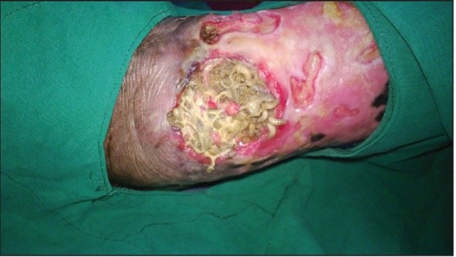
Figure 1: Wound Myiasis
DISCUSSION
Female flies deliberately lay eggs in living organisms for the purpose of the development of their young. Living organisms provide a food source for the developing maggots. They can lay 50-300 eggs at a time. These eggs hatch about 8-12 hours later and attain full growth in about 2-3 days (1,2). Multiple types of myiasis (infection with fly larvae) can occur. Cutaneous myiasis occurs when a fly digs under the skin to lay its eggs. This creates a small nodule or lesion resembling an insect bite or pimple. There is usually a small pore in the center for the maggot to breathe. Another type of myiasis is creeping myiasis, which is when the maggot moves through the body to find a place for development. This occurs when the patient becomes an accidental, but not suitable, host for the maggot. Unfortunately, this movement of the maggot causes a painful “creeping” sensation for the patient. Wound myiasis is when the maggot lays eggs in decaying or pus-filled flesh. Accidental myiasis is the accidental ingestion of maggots or eggs, which results in nausea, vomiting, and gastrointestinal upset. Body cavity myiasis is when maggots invade the eyes, nose, mouth, or ears [1]. Myiasis is more common in warmer climates and tropical countries, as well as rural regions. Other risk factors include diabetes, peripheral artery disease, neurologic disorders, poor personal hygiene, homelessness, or substance abuse. Common sites for myiasis include the head, neck, trunk, and lower limbs [3].
MANAGEMENT
This patient had wound myiasis requiring removal of the maggots before admission for treatment of his necrotic ulcers and surrounding cellulitis. Management included two steps. First, the maggots were removed. Second, the maggots were contained and properly disposed of, so as to not cause a secondary infestation within the hospital. There are minimal documented case reports detailing the removal of maggots from wounds and outlining the best approach. Elder et al. proposes that a safe technique for removal is to use the yankauer and suction. They propose that the clinician should first suction off the superficial maggots, which would then allow the deeper maggots to emerge. The maggots would collect in the suction bin to be safely disposed of in a contained manner [4]. Other proposed methods include pouring sterile water over the maggots and collecting them below the stream of water, soaking the limb in sterile water so that the maggots fall off, or removing the maggots with forceps [5].
All the above methods were attempted but failed to remove the maggots from the patient in this case. When the yankauer technique was attempted, the maggots became stuck in the tubing, never making it to the canister, and blocking any further suction power. Both of the sterile water methods did not work because the maggots seemed to hang on and be unhindered by the water. Moreover, the ones that were submerged simply crawled to dry tissue. Lastly, the idea of removing several hundred maggots one by one with forceps in a busy emergency department was unrealistic. Ultimately, the physicians in this case found that pouring dilute hydrogen peroxide over the maggots and then gently wiping the the area with gauze was the best solution. This allowed for the maggots to become stunned and stop burrowing into the tissue long enough to be easily removed in groups. Once the outer layer of maggots was removed, the deeper ones emerged from the ulcers, and the aforementioned steps of the procedure were repeated. The maggots were then be wiped into a bin to be collected. The clinican then poured solidifier into the container, which trapped the maggots into the solution. They were then placed in a sealed container, then into a closed bag, and, ultimately, properly disposed of without causing an infestation. Note: This protocol may differ at different facilities but is what was recommended by infection control at the institution at which this patient was treated.
CONSLUSION & TAKE-AWAYS
1. There are many techniques for maggot removal in the emergency department.
2. In this case, the team found that stunning the maggots with hydrogen peroxide prior to wiping them off of the wound was the most effective method.
3. Check with your hospital's infection control to find the protocol for safe disposal.
Author: Giovanna Deluca, MD is a fourth-year emergency medicine resident at Rhode Island Hospital/Brown University
Faculty Reviewer: Kristina McAteer, MD is an emergency medicine attending at Rhode Island Hospital/Newport Hospital
References
1. Sunny B, Sulthana L, James A, Sivakumar T. Maggot Infestation: Various Treatment Modalities. J Am Coll Clin Wound Spec. 2018 Mar 30;8(1-3):51-53. doi: 10.1016/j.jccw.2018.03.002. PMID: 30276127; PMCID: PMC6161638.
2. Spradbery J.P. Screw-worm fly: a tale of two species. Agric Zool Rev. 1994;6:1–62.
3. Andreatta, E., Bonavina, L. Wound myiasis in Western Europe: prevalence and risk factors in a changing climate scenario. Eur Surg (2021). https://doi.org/10.1007/s10353-021-00730-y
4. Elder JW, Grover CA. Wound debridement: lessons learned of when and how to remove "wild" maggots. J Emerg Med. 2013 Oct;45(4):585-7. doi: 10.1016/j.jemermed.2013.01.022. Epub 2013 Apr 25. PMID: 23623148.
5. BCProvincial NursingSkin& WoundCommittee in collaboration with NSWOCs/WoundClinicians. (2019, July). Procedure: Removal of non-therapeutic maggots (myiasis) - CLWK. British Columbia Provincial Nursing Skin & Wound Committee Procedure: Removal of Non-therapeutic Maggots (Myiasis). Retrieved September 9, 2022, from https://www.clwk.ca/get-resource/removal-of-non-therapeutic-maggots-myiasis-procedure/
6. Photo: https://woundcareadvisor.com/using-maggots-in-wound-care-part-1-vol3-no4/