When laughter isn’t the best medicine: The serratus anterior plane block for multiple rib fractures
Case
A 67-year-old man presents to the emergency department after a mechanical fall from standing. During his trauma evaluation, he is found to have severe right-sided chest wall tenderness. His imaging demonstrates multiple right-sided rib fractures with an otherwise unremarkable workup. The patient is in significant discomfort and is given acetaminophen and morphine. Despite this, he remains in severe pain and has difficulty taking deep breaths. The patient is given an incentive spirometer and his maximum vital capacity is measured at 500 ml. What complications is this patient at risk of developing? What interventions can be performed in the emergency department to help manage his pain?
DISCUSSION
Introduction
Rib fractures are one of the most common traumatic injuries seen in the emergency department. Severe pain from multiple rib fractures can impair respiratory function and decrease the ability to clear secretions, leading to an increased risk of atelectasis, pneumonia and ARDS [1]. For patients with multiple rib fractures, morbidity increases significantly for patients over 45 years of age and mortality increases 5-fold for patients over 65 years of age [2,3]. Early and aggressive pain control is critical to decrease these complications. Medications commonly used to treat pain from rib fractures include acetaminophen, NSAIDs, and opioids. However, this combination may not provide adequate analgesia in all patients [1]. In addition to respiratory complications, poorly controlled pain is associated with increased risk of delirium in the elderly. Opioid pain medications can also cause respiratory depression, impaired cough reflex, and delirium [4]. As a result, regional blocks are being used more frequently in the emergency department as a safe and effective pain management adjunct in patients with multiple rib fractures.
Evidence
The serratus anterior plane block (SAPB) is a plane block which can be used to provide regional anesthesia for a variety of indications, including rib fractures and tube thoracostomy placement. Patients with multiple rib fractures who underwent SAPB performed by trained ED physicians experienced a significant reduction in pain scores and increase in maximum vital capacity (5,6). While no studies directly compare the effectiveness of the SAPB based on rib fracture location, it is believed to provide better analgesia for anterolateral rib fractures compared to posterior rib fractures (7).
Anatomy
The lateral cutaneous branches of the thoracic intercostal nerves provide the sensory innervation of the chest wall. The thoracic intercostal nerves, which originate from the spinal thoracic nerves, run anterolaterally alongside the intercostal artery and vein in the costal groove on the inferior rib margins. At the midaxillary line, the lateral cutaneous branches exit through the intercostal and serratus anterior muscle. They continue to travel anteriorly in the fascial plane just superficial to the serratus anterior and deep to the latissimus dorsi, providing sensory and motor innervation to the chest wall (Figure 1). This fascial plane is the target of the serratus anterior plane block. When performed correctly, the block should provide anesthesia to the T2-T9 dermatomes of the anterolateral chest wall (Figure 2).

Figure 1: Anatomy and innervation of the chest wall [8]

Figure 2: Distribution of analgesia from serratus anterior plane block [9]
Supplies
High-frequency linear transducer
Sterile probe cover
Sterile gel
Chlorhexidine or betadine solution
Sterile gloves
1% lidocaine with epinephrine in syringe with 27-gauge needle (for skin wheel)
0.5% bupivacaine in 30 ml syringe (see Figure 3 for alternative agents and dosing guidelines). If max dose is under 30 ml, dilute anesthetic further with sterile saline to reach a total of 30 ml of anesthetic/saline mixture
Peripheral nerve block needle with extension tubing
a. Alternatively, 22g spinal needle with IV extension tubing
Intralipid (must be present in the department prior to any performing nerve block)

Figure 3: Dosing guidelines for commonly used local anesthetics [10]
The Procedure
Position the patient in either the supine or the lateral decubitus position with the affected side facing up. Identify the mid-axillary line and place the transducer at the level of the fifth rib in the transverse plane (Figure 4). Identify the fifth rib, pleura, intercostal muscles, serratus anterior, latissimus dorsi and anterior fascial plane just superficial to the serratus anterior. This is the target of the block.

Figure 4: Ultrasound image showing target fascial plane and anatomy of the chest wall, as well as appropriate probe positioning [8]
Prep and drape the patient in the usual sterile fashion. Prep the needle and tubing with a saline flush. Draw up the local anesthetic in a 30 ml syringe. If necessary, draw up additional saline into the syringe to reach a total volume of 30 ml mixture of local anesthetic and saline. Place a sterile probe cover over the probe and place sterile gel onto the field.
Obtain the previous view using the linear transducer. Place a small amount of local anesthetic (typically 1% lidocaine with epinephrine) and make a skin wheel at the insertion site. Insert the needle in-plane with the ultrasound probe and keep the needle tip in view. Be sure you are aware of exactly where the needle tip is which is easier to do with an in-plane technique. Always keep the needle tip directed at the rib, as this will help decrease the risk of accidental pleural puncture and subsequent pneumothorax. Continue advancing the needle until it enters through the fascial plane just superficial to the serratus anterior. At this point, it is helpful to have a second clinician available to hold the syringe and draw back, and then push the anesthetic, while the proceduralist holds the needle and ultrasound probe. Hydrodissect with a small amount of saline to be sure your needle tip is in the correct place. Once the needle tip location is confirmed, slowly inject 30 ml of saline/anesthetic mixture into this plane. A collection of anechoic fluid will begin to form in this plane (Figure 5).

Figure 5: Ultrasound image demonstrating anterior fascial plane and anechoic collection of local anesthetic [8]
While there is limited evidence, some recommend targeting the fascial plane just deep to the serratus anterior may improve the analgesic effect for posterior rib fractures [7]. Comparison of the anterior and posterior approach can be seen in Figure 6. Remove the needle and place a dressing on the puncture site. Continue to monitor the patient after completion of the procedure to determine whether the block was effective and to identify any complications.
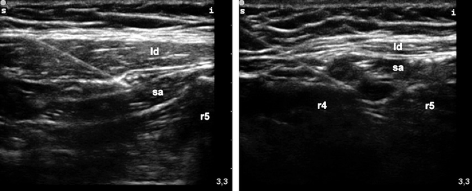
Figure 6: Comparison of needle tip placement for anterior (left) versus posterior (right) serratus anterior plane block. Ld = latissimus dorsi, sa = serratus anterior, r4 = rib 4, r5= rib 5. [11]
Complications
Local anesthetic systemic toxicity (LAST) is a severe complication when performing any nerve block. Neurological symptoms include metallic taste, tinnitus, perioral numbness, dizziness, vision changes or seizure, while cardiac complications include decreased contractility, refractory hypotension, heart block, arrhythmias, or cardiac arrest. The treatment of LAST is intravenous lipid emulsion, often known as intralipid, and this should be confirmed to be present in the emergency department prior to any nerve block. Pneumothorax is a rare complication. However, the risk of this complication can be decreased by keeping the needle tip visualized and directed at the rib at all times, as described above. Additional complications include nerve injury, vessel injury, and rebound pain.
TAKE-AWAYS
● Multiple rib fractures carry significant morbidity and mortality in middle-aged and elderly patients, primarily due to respiratory driven complications
● The SAPB is an effective tool to decrease pain and improve vital capacity in patients with multiple rib fractures
● The SAPB can also be used as pre-procedure block for chest tubes or axillary abscess drainage
● The effectiveness of the SAPB may be limited in posterior rib fractures
● Recognize the signs, symptoms and management of LAST
AUTHOR: Brendan Holmes, MD is a first-year emergency medicine resident at Brown University/Rhode Island Hospital.
FACULTY REVIEWER: Kristin Dwyer, MD is Assistant Professor of Emergency Medicine at Warren Alpert Medical School of Brown University and Emergency Ultrasound Fellowship Director
REFERENCES
Kim M, Moore JE. Chest Trauma: Current Recommendations for Rib Fractures, Pneumothorax, and Other Injuries. Curr Anesthesiol Rep. 2020;10(1):61-68. doi:10.1007/s40140-020-00374-w
Holcomb JB, McMullin NR, Kozar RA, Lygas MH, Moore FA. Morbidity from rib fractures increases after age 45. J Am Coll Surg. 2003;196(4):549-555. doi:10.1016/S1072-7515(02)01894-X
Bergeron E, Lavoie A, Clas D, et al. Elderly trauma patients with rib fractures are at greater risk of death and pneumonia. J Trauma. 2003;54(3):478-485. doi:10.1097/01.TA.0000037095.83469.4C
Clegg A, Young JB. Which medications to avoid in people at risk of delirium: a systematic review. Age Ageing. 2011;40(1):23-29. doi:10.1093/ageing/afq140
Kring RM, Mackenzie DC, Wilson CN, Rappold JF, Strout TD, Croft PE. Ultrasound-Guided Serratus Anterior Plane Block (SAPB) Improves Pain Control in Patients With Rib Fractures [published online ahead of print, 2022 Feb 2]. J Ultrasound Med. 2022;10.1002/jum.15953. doi:10.1002/jum.15953
Paul S, Bhoi SK, Sinha TP, Kumar G. Ultrasound-Guided Serratus Anterior Plane Block for Rib Fracture-Associated Pain Management in Emergency Department. J Emerg Trauma Shock. 2020;13(3):208-212. doi:10.4103/JETS.JETS_155_19
Almeida CR. Serratus anterior plane block for posterior rib fractures: why and when may it work?. Reg Anesth Pain Med. 2021;46(9):835-836. doi:10.1136/rapm-2020-102098
Nagdev A, Mantuani D, Durant E, Herring A. Ultrasound-guided Serratus Anterior Plane Block can help avoid opioid use for patients with rib fractures - page 2 of 4. ACEP Now. https://www.acepnow.com/article/ultrasound-guided-serratus-anterior-plane-block-can-help-avoid-opioid-use-patients-rib-fractures/2/. Published April 5, 2019. Accessed February 7, 2022.
Avila J. 5 minute sono – serratus anterior block. Core Ultrasound. https://www.coreultrasound.com/serratus/. Published June 19, 2020. Accessed February 7, 2022.
Block Basics: What med? Highland EM Ultrasound. http://highlandultrasound.com/med-guide. Accessed February 7, 2022.
Blanco R, Parras T, McDonnell JG, Prats-Galino A. Serratus plane block: a novel ultrasound-guided thoracic wall nerve block. Anaesthesia. 2013;68(11):1107-1113. doi:10.1111/anae.12344