Global Emergency Medicine: An unusual case of epistaxis

“Myiasis infestations are classified either by the host-parasite relationship or by the location of the larvae, with cutaneous myiasis being the most common form.”
The Case:
An 84-year-old female with no significant medical history presents to an emergency department in rural, far-west, Nepal with three days of nasal congestion, discomfort, and intermittent bleeding. She reports foul smelling nasal discharge. She denies fevers, vomiting, diarrhea, shortness of breath, or previous similar symptoms. She has had no recent trauma and is not anti-coagulated.
Vitals:
BP 150/10 | Pulse 102 | Temp 37.4°C (Temporal) | Resp 16 | SpO2 94%
Physical Exam:
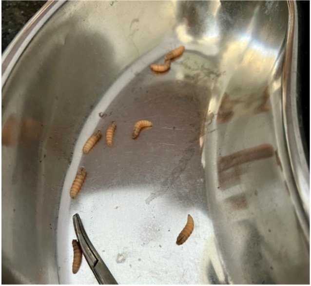
You note an uncomfortable-appearing elderly female. She has dried blood within each nare and at their bases, with more blood on the left. See the image below. During an intranasal inspection, you notice several small white objects moving within the nasal cavity. The patient blows her nose and multiple larvae are released from her nose.

Figure 1: the patient’s nose
Diagnosis:
Intranasal Myiasis
Discussion:
Myiasis (myia: Greek for fly) is the infestation of fly larvae (maggots) in human or other vertebrate animal tissue. [1] Although the type of fly varies by region, the two most common flies leading to human myiasis infestations are Dermatobia hominis (human botfly) and Cordylobia anthropophaga (tumbu fly). Infestations occur when the adult fly lays eggs directly onto human tissue, or they lay eggs on a vector who transmit them to humans when taking a blood meal. [1] Myiasis infestations are classified either by the host-parasite relationship or by the location of the larvae, with cutaneous myiasis being the most common form. [2] The larvae survive on the human host by feeding on human tissue or liquid body substance. Rarely encountered in the United States, myiasis is more commonly found in tropical and subtropical regions, especially in rural locations. [3] Myiasis infections often occur in patients who are ill from another disease or who are incapacitated. During the patient’s evaluation, she was found to have consistently elevated blood glucose levels and was diagnosed with diabetes, one of the major risk factors for developing myiasis infestations. This patient also resided in a rural setting with proximty to livestock, another risk factor. [4] Many myiasis infestations are self-limiting—once the larvae emerge, the lifecycle is broken, and the wounds will heal. However, secondary infections such as cellulitis are the most common complications. [5] Ophthalmomyiasis and nasal myiasis have the additional risk of the larvae burrowing into brain matter, leading to meningitis, pneumocephalus, and neurological sequela. [6,7] The most common symptoms associated with nasal myiasis are epistaxis, foul smell, the passage of worms, facial pain, nasal obstruction, nasal discharge, headache, dysphagia, and foreign body sensation in the nose. [8]

https://www.cdc.gov/parasites/myiasis/biology.html
Treatment of myiasis:
There are four main approaches to myiasis treatment depending on the type of infestation and available resources: debridement/irrigation, occlusion/suffocation, surgical removal, and systemic/topical ivermectin. Debridement and irrigation are best used for wound myiasis. The occlusion/suffocation technique is performed by placing petroleum jelly, liquid paraffin, beeswax, or heavy oil over the punctate opening, which leads to the suffocation of the larvae. The larvar will then spontaneously emerge in search of oxygen. When using this approach, allowing the larvae to evacuate spontaneously is essential. If they asphyxiate while still within the tissue, local reaction and infection risk will require surgical intervention. [8] Deep cutaneous myiasis infestations or infestations in orifices close to the brain often require surgical removal when available. Advanced imaging, such as CT and MRI, or direct visualization using a nasal endoscope, have been recommended for nasal myiasis to determine the extent of the infestation. However, these diagnostic and therapeutic measures may not be feasible in limited resource settings. When invasive surgical debridement is unavailable for nasal myiasis, it has been proposed that packing the nose with a chloroform and turpentine (1:4) mixture followed by manual removal of the dead maggots is an alternative treatment protocol. [9]
Systemic ivermectin, oral or IV, has been proposed as a primary and neoadjuvant treatment for cavitary myiasis. [6] Larvae utilize their hook-like structures to bury into the tissue to avoid removal. Thus, when used as an adjunct treatment before surgical debridement, ivermectin has been shown to reduce tissue destruction during surgical extraction. [10,11]
Additionally, tetanus vaccination status should be updated if not current, as myiasis infestation can lead to infection with Clostridium tetani. [6]
Case Resolution:
Given the available resources the patient was treated with a combination of oral ivermectin 200 μg/kg and albendazole 400 mg, followed by a second dose of ivermectin. To treat likely secondary intranasal soft tissue infection, she was started on ceftriaxone 1 gm twice daily. She was monitored for five days, during her hospitalization numerous larvae were spontaneously evacuated and her symptoms resolved, allowing her to be discharged home.
Authors: Derek Lubetkin, MD is a recent graduate of the Brown Emergency Medicine residency program & Niranjan Rijal, MBBS
Faculty Reviewer: Ramu Kharel, MD, MPH, CTropMed®, is an Assistant Professor of Emergency Medicine at Warren Alpert Medical School of Brown University and the Nepal Cluster Coordinator for the Saxena Center for Contemporary South Asia
References:
1. https://www.cdc.gov/parasites/myiasis/index.html
2. Hall M, Wall R. Myiasis of humans and domes- tic animals. Adv Parasitol 1995;35:257–334.
3. Zumpt F. Myiasis in Man and Animals in the Old World. London: Butterworths; 1965.
4. Auerbach PS. Arthropod envenomation and parasitism. Wilderness Medicine. 5th ed. Philadelphia, PA: Mosby Elsevier; 2007. 969-974.
5. Davis RF, Johnston GA, Sladden MJ. Recognition and management of common ectoparasitic diseases in travelers. Am J Clin Dermatol. 2009. 10(1):1-8.
6. Bolognia JL, Jorizzo JL, Rapini R. Cutaneous myiasis. Dermatology. 2nd ed. Mosby Elsevier; 2008. Vol 1: 1300-01.
7. Kuruvilla G, Albert RR, Job A, Ranjith VT, Selvakumar P. Pneumocephalus: a rare complication of nasal myiasis. Am J Otolaryngol. 2006 Mar-Apr;27(2):133-5.
8. Maier H, Hönigsmann H. Furuncular myiasis caused by Dermatobia hominis, the human botfly. J Am Acad Dermatol. 2004 Feb. 50(2 Suppl):S26-30.
9. Sharma H, Dayal D, Agrawal SP. Nasal myiasis: review of 10 years experience. J Laryngol Otol. 1989 May. 103(5):489-91.
10. Tsuda S, Nagaji J, Kurose K, Miyasato M, Sasai Y, Yoneda Y. Furuncular cutaneous myiasis caused by Dermatobia hominis larvae following travel to Brazil. Int J Dermatol.
11. Osorio J, Moncada L, Molano A, et al. Role of ivermectin in the treatment of severe orbital myiasis due to Cochliomyia hominivorax. Clin Infect Dis. 2006 Sep 15. 43(6):e57-9