A Toddler’s Tibia: Common Yet Obscure
CASE
A 3-year-old male presents to the pediatric emergency department with leg pain. Prior to arrival, the patient had been at daycare and was found by staff crying, reporting pain to his right leg. Daycare staff denied any trauma and noted that the child had been playing happily throughout the morning. His parents were understandably concerned and the patient’s father reported that this had been his fourth visit to the emergency department for fractures since he started walking at 12 months of age. The patient was otherwise healthy. Additionally, the parents had no concerns about safety at the daycare, as the patient’s sibling attended the same center without any issues. The daycare center also had “nanny cams” and provided videos to the parents showing no specific injury or mistreatment.
Vital signs were normal. On physical exam, the chid was sitting comfortably, playing with a toy, but refusing to move his right leg or foot. He had a small area of ecchymosis to the anterior right shin without obvious deformity. The right leg was neurovascularly intact. No other bruising or injuries were appreciated. An X-ray of the leg was obtained. (Figure 1)
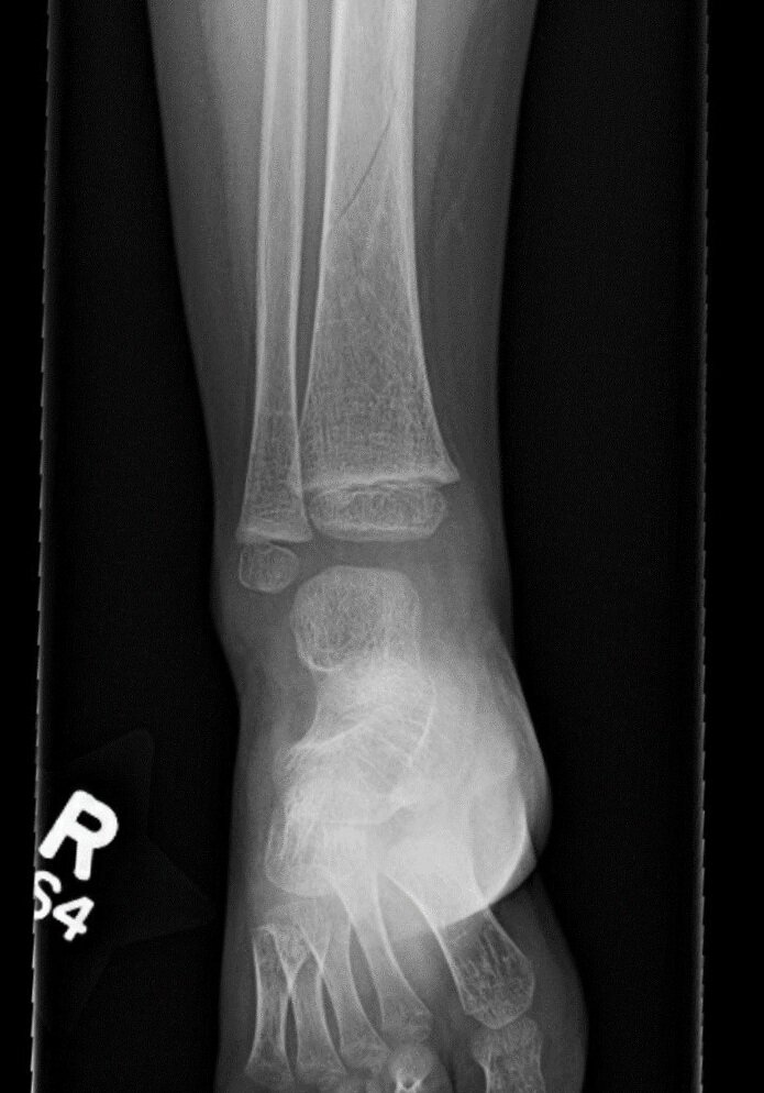
Figure 1. Spiral fracture of the right distal tibia
DIAGNOSIS
Non-displaced spiral fracture of the tibia, also known as a “toddler’s fracture”
DISCUSSION
A brief history lesson
In a March 1964 address to the Canadian Association of Radiologists, Dr. J.S. Dunbar, MD stated that when a child "fails to bear weight on the leg," he most often has an obscure fracture of the tibia called the "toddler's fracture". [1]

Typical presentation
Toddler’s fractures usually occur after a child begins to ambulate on two legs, thus placing weight on the tibia. The incidence is greatest between the ages of 9 months and 3 years. The child typically presents with refusal to bear weight and increased irritability. There may or may not be a report of antecedent trauma or injury, and often the injury may be seemingly insignificant. [1,2]
Radiology
A toddler’s fracture is a minimally displaced or non-displaced spiral fracture, usually of the tibia, typically encountered in toddlers. (Figure 1) Importantly, this definition does not include spiral femoral fractures, which should raise suspicion of non-accidental trauma (NAT). [3] When a toddler’s fracture is suspected, AP and lateral views of the tibia and fibula should be obtained. (Figure 2)
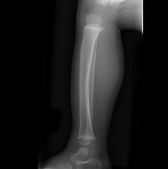
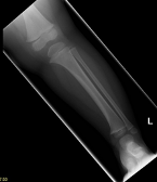
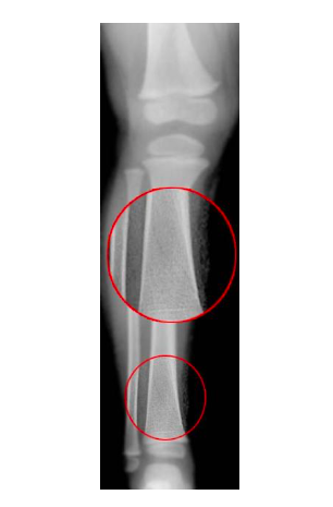
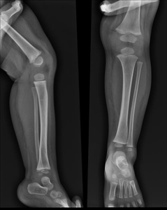
Figure 2. Examples of AP and lateral views of tibial spiral fractures
Management
Historically, toddler’s fractures are managed non-operatively by immobilization with a long leg cast with orthopedic follow-up. More recent studies, however, have shown that a short leg cast or CAM boot has similar outcomes. [2,4] Plain films may be negative on initial evaluation, however, if injury and presentation are consistent with toddler’s fracture, a CAM boot/short leg splint or cast should be considered and follow-up radiographs should be obtained seven days later. Orthopedic follow-up should occur within 7-10 days of injury. Most children will return to ambulation after 3-4 weeks. Complications are rare, as the injury often has a low-energy mechanism and young patients heal quickly. [5]
Expanding the differential
Toddler’s fractures are generally straightforward and are not traditionally associated with abuse. Nonetheless, with any child presenting with a fracture, it is important to consider if the injury may be due to NAT, as an estimated 1-10% of pediatric injuries seen in the ED are due to abuse. [6] In our case, the three previous fractures fit the pattern of age-expected fractures. There was also documentation that parents presented immediately after injuries and that these were isolated injuries without other signs of NAT.
Additionally, a good medical history, including any previous fractures, should be obtained to assess for the possibility of more rare presentations, such as metabolic or connective tissue disorders. Of note, on this patient’s physical exam, he was noted to have pronounced blue sclera, one of the characteristic features of osteogenesis imperfecta (OI) Type I. This collagen disorder is the mildest form of OI and fractures tend to begin when a child starts to walk. [7] Although fractures usually decrease after puberty, long-term sequelae may include shorter stature and early hearing loss. Patients with OI have blue-appearing sclera due to defective collagen, resulting in a thinner covering of the eye revealing the choroidal veins. Although not often a diagnosis that will be made in the emergency department, suspicion for OI should be discussed with the parents, and a referral to genetic counseling may be indicated.
CASE RESOLUTION
The patient was placed in a long leg cast, made non-weight-bearing on the right leg, and discharged home with orthopedic follow-up and a genetic counseling referral.
TAKE-AWAYS
A toddler’s fracture is a minimally or non-displaced spiral fracture of the tibia that occurs between 9 months and 3 years of age
The patient or parents may or may not report antecedent trauma or injury
Traditionally, immobilization in a long leg cast has been the treatment of choice for toddler’s fractures, but new data suggests a short leg cast or CAM boot may be just as effective
Plain films may be negative on initial evaluation, thus if injury and presentation are consistent with toddler’s fracture, a CAM boot/short leg splint or cast should be considered until follow-up films can be obtained seven days later
Patients should be referred to an orthopedist for follow up in 7-10 days
Author: Derek Lubetkin, MD is a second-year Emergency Medicine resident at Brown University/Rhode Island Hospital.
Faculty Reviewer: Jeffrey Feden, MD
References
1. Dunbar, JS et al. A Common, Obscure Fracture of the Tibia -- The Toddler's Fracture. J Can Assoc Radiol. 1964;15:136.
2. Bauer JM & Lovejoy SA. Toddler's Fractures: Time to Weight-bear With Regard to Immobilization Type and Radiographic Monitoring. J Pediatr Orthop. 2019;39(6):314.
3. Toddler Fracture [Internet]. Rasuli, B & Gaillard, F, Radiopaedia.org [Accessed on 2020 April 12]. Available from: https://radiopaedia.org/articles/toddler-fracture?lang=us
4. Leffler LC, Tanner SL, Beckish ML. Immobilization Versus Observation in Children With Toddler's Fractures: A Retrospective Review. J Surg Orthop Adv. Summer;27(2):142.
5. Mashru, RP et al. Tibial shaft fractures in children and adolescents. J Am Acad Orthop Surg. 2005 Sep; 13(5):345-52.
6. Narang, et al. Legal Issues in Child Maltreatment Pediatr Clin North America 2014; 1049–1058.
7. Rauch F & Glorieux FH. Osteogenesis imperfecta. Lancet. 2004;363(9418):1377-1385.
Image Credit
Toddler Fracture [Internet]. Rasuli, B & Gaillard, F, Radiopaedia.org [Accessed on 2020 April 12]. Available from: https://radiopaedia.org/articles/toddler-fracture?lang=us