

…Patients who underwent POCUS examination for identification of an IUP had a length of stay in the emergency department that was over an hour shorter than those who went for comprehensive ultrasound. Emergency physician proficiency in POCUS in early pregnancy could improve patient flow and satisfaction…

This patient is a 57 year old female who had no known medical history who presented to the emergency department for visual disturbances…

Point-of-Care Ultrasound (POCUS) is a diagnostic imaging technique often used at the bedside in a variety of settings including emergency care, cardiology, intensive care, anesthesiology and obstetrics among others. Alongside the rapid evolution of machine learning (ML) and deep learning (DL) algorithms in the health informatics space, “intelligent” POCUS systems are concurrently transforming. From guiding novice trainees to automating clinical calculations, these algorithms are making ultrasound faster, more accurate, and more accessible…

25-year-old male with history of nephrolithiasis (prior right UVJ stone, 4x6mm visualized on CT a year ago) presenting with acute onset severe L sided flank pain that woke him from sleep...

A previously healthy ten-year-old girl presented to the pediatric emergency department, accompanied by her mother, with two days of lower abdominal pain and urinary frequency and urgency...

A 75-year-old-female with a past medical history of dementia, diaphragm dysfunction, OSA on CPAP, presents to the Emergency Department for a fall. She is not on any blood thinning medications. Patient was found in the basement by her husband who believes she tripped over her oxygen tubing…

Necrotizing fasciitis is a life-threatening soft tissue infection that causes necrosis of the muscle fascia and subcutaneous tissues…

Calciphylaxis is a life-threatening vasculopathy that results from the deposition of calcium in the arteriolar vasculature of the deep dermis and subcutaneous adipose tissue, which causes occlusion of the affected blood vessels and overlying tissues and skin…

A 41-year-old G1P0 female at 7 weeks gestation presents to the emergency department (ED) with 3 days of mild lower abdominal cramping and intermittent vaginal spotting. Nothing makes the cramps better or worse and the spotting randomly occurs throughout the day. She rates the cramping at a 7/10 and describes it as “bad period cramps.” She has no other symptoms and is nervous that “something is wrong with the baby”…

A 68-year-old male presents to the emergency department (ED) with one week of worsening shortness of breath. He has no known cardiac history and has rarely seen a physician. He reports shortness of breath with exertion, mild non-productive cough, and subjective fever. He is not having chest pain. He is a long-time smoker but denies other drugs. Vitals are as follows: heart rate (HR) of 102, blood pressure (BP) of 105/70, afebrile, 89% on room air (RA), respiratory rate of 20. On exam the patient exhibits labored breathing, crackles in bilateral lower lung fields and +1 pitting edema in bilateral lower extremities…

A 31 y.o. male presents to the Emergency Department with a chief complaint of swelling of his right leg. He has no past medical history, and his social history is significant for polysubstance use disorder and housing insecurity. He primarily injects opioids and cocaine and has had periods of time where he received medication assisted treatment with buprenorphine. On exam, there is an area of erythema and swelling on the lower right extremity that does not overly the joints. The area is warm and tender to palpation. There is no obvious wound or purulent drainage. The remainder of a 12 point Review of Systems is negative…

A 14-year-old female presented to the emergency department (ED) with persistent left upper quadrant (LUQ) pain. She was last seen in the ED one week prior for similar symptoms. Initially, she was diagnosed with a urinary tract infection (UTI), treated with IV ceftriaxone, and discharged home on a course of cefdinir. On re-presentation, she complained of persistent LUQ pain, fevers with a Tmax of 103 F, and one episode of emesis overnight. Physical examination revealed abdominal tenderness in the LUQ, without ipsilateral CVA tenderness…

A 53-year-old male with hypertension presents to the emergency room with painful swelling of the right first toe at the MTP joint. The patient was in good health until a few days ago, where he was attending a college reunion. The swelling and pain increased overnight. The patient is having difficulty walking this morning due to the pain. He tried taking two Advil for the pain, which helped bring the pain from an 8/10 to a 6/10…

A 67-year-old man presents to the emergency department after a mechanical fall from standing. During his trauma evaluation, he is found to have severe right-sided chest wall tenderness. His imaging demonstrates multiple right-sided rib fractures with an otherwise unremarkable workup. The patient is in significant discomfort and is given acetaminophen and morphine. Despite this, he remains in severe pain and has difficulty taking deep breaths. The patient is given an incentive spirometer and his maximum vital capacity is measured at 500 ml. What complications is this patient at risk of developing? What interventions can be performed in the emergency department to help manage his pain?

An 83-year-old male with a history of HFrEF (40%), CAD, hypertension, and hyperlipidemia presented to the ED with generalized weakness and fatigue. His vitals were remarkable for hypotension with a MAP of 58. On physical exam, the patient appeared non-toxic and was able to answer questions appropriately. He did not appear frankly fluid overloaded, other than mild, chronic peripheral edema. Additional laboratory studies and chest x-ray were performed. A fluid bolus was given to treat his hypotension, however there was minimal response. Given his heart failure, there was concern that aggressive fluid resuscitation would result in pulmonary edema. Additionally, inserting a central line and starting vasopressors is not without risk. Fortunately, this clinical team knows how to use ultrasound to do a proper volume assessment to manage this patient appropriately…

“It would be unusual to go too many shifts as an emergency medicine provider without seeing the classic elderly female patient with hip pain after a fall. On exam, the patient would likely be uncomfortable with their hip appearing shortened and externally rotated on exam. Before you even view the X-ray, you know they likely fractured their hip. You ascertain whether it was a mechanical fall, if there were other injuries, if the injury is open or closed and if the patient is neurovascularly intact. You proceed to order your imaging and consider your plan for pain control. Sound familiar…”

Renal ultrasound can be important in determining diagnosis, management, and next steps in the evaluation of a patient with flank or abdominal pain, hematuria, concern for stone and more.

A 20-year-old female presents to the emergency department (ED) with a chief complaint of a headache. Her headache started 8 days ago and is described as bifrontal. It is positional and gets worse when she bends forward but improves when she is sitting or standing. She had presented to 3 other EDs prior to this visit for her symptoms, and each time she was discharged with an intractable headache after receiving a migraine cocktail to no effect…

…3 days of floaters and “lightning bolt” white flashes in the temporal field of her right eye in the absence of eye trauma. She also reported right periorbital pain. She did not have blurry vision, diplopia, loss of vision, scalp tenderness, jaw claudication, fevers, chills, or weight loss. She denied photophobia despite wearing sunglasses in the Emergency Department. Her mother had died from choroidal melanoma, but the patient had not seen an ophthalmologist in 15 years.
Her uncorrected visual acuities were 20/60 OD and 20/40 OS. Intraocular pressure, pupillary reaction, and extraocular movements were normal. Point of care ultrasound (POCUS) revealed three hyperechoic, solid lesions with posterior acoustic shadowing in her right eye….

…complained of significant superior lateral knee pain and swelling, as well as difficulty moving his leg due to pain. On exam, he had notable swelling and tenderness of the left knee, particularly along the superior aspect of the patella. After controlling his pain with intravenous medications, he was able to flex at the hip but was unable to extend his lower leg at the knee or to perform a straight leg raise….

…The differential diagnosis included acute coronary syndrome, CHF exacerbation, pneumonia, and cardiac amyloidosis.
EKG showed sinus tachycardia with low voltage. Laboratory evaluation was unremarkable, with a negative troponin and high normal BNP. Chest radiograph showed bilateral infiltrates concerning for mild pulmonary congestion.
POCUS was obtained….

An octogenarian gentleman with a history of atrial fibrillation, congestive heart failure, chronic kidney disease, and insulin-dependent diabetes presented to the emergency department with three weeks of pain and swelling near the right side of his face. He awoke that morning with a new symptom -- difficulty swallowing…
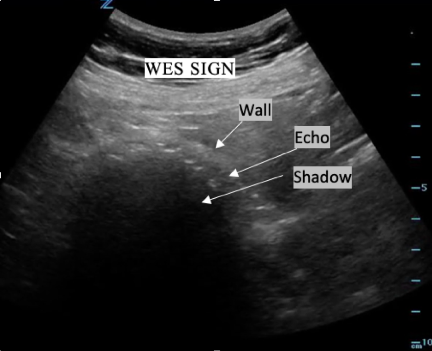
40 year old female with normal vital signs and a chief complaint of right upper quadrant abdominal pain associated with nausea and vomiting. She has never had this pain before. She has no past medical and no past surgical history. She denies vaginal and urinary symptoms. She undergoes right upper quadrant ultrasound which confirms the diagnosis…

“A 42 year old male with a past medical history of paroxysmal atrial fibrillation and L4 radiculopathy due to a remote back injury presents to the ED with chief complaint of right foot and calf pain. The pain is described as cramping and burning that gradually worsened over the 3 days prior to presentation. He is not on anticoagulation. He denies…”

Nasogastric tube (NGT) placement is a commonly performed procedure in the emergency department (ED) that is indicated for a variety of diagnostic and therapeutic purposes…

A 53-year-old woman with a past medical history of hypertension arrived to the Emergency Department with left arm pain shortly after a mechanical fall on an outstretched hand…

A 53-year-old male with a PMH of GERD presents to the emergency department with eye pain, photophobia, and vision loss after getting popped in the eye by a broken bungee cord. He believes the tip of the cord came around “like a whip” and struck him directly on the eye…

She goes to see the patient, a 27 year old male with no past medical history who spent the afternoon surfing. He was walking back to his car barefoot and stepped on something that made him jump. He has no idea what it could be but “it’s still in there doc!” …

A 24-year-old female with a past medical history of polycystic ovarian syndrome and urinary tract infections, who presented to the emergency department (ED) via urgent care with acute on chronic worsening abdominal pain and a cystic structure in the pelvis on computed tomography (CT) scan…
Pearls and pitfalls: Scan in BOTH longitudinal and transverse planes. To avoid rib shadows, oblique the probe to align with the rib space rather than holding the probe straight up and down. Slowly fan through the kidney in its entirety to evaluate for hydronephrosis and for the presence of a kidney stone…